Seven images were shortlisted for this year’s annual Science and Medical Image competition, showcasing the eye-catching science being carried out at the ICR. Three winners were selected by a judging panel and the fourth was chosen by the public based on votes on social media.
Images of cancer research
Our annual imaging competition celebrates the incredible research by our scientists at the ICR and The Royal Marsden, and the arresting images they produce – often they are capturing cellular processes in action, which is key to making new discoveries or deepening our understanding of clinical problems affecting cancer patients.
The selection chosen for the judges’ shortlist covers different cancer types and different scientific techniques such as immunofluorescence, microscopy and 3D printing.
Judges’ winner
Dr Maria Taskinen, Postdoctoral Training Fellow in the Signal Transduction and Molecular Pharmacology Group in the Division of Cancer Therapeutics.
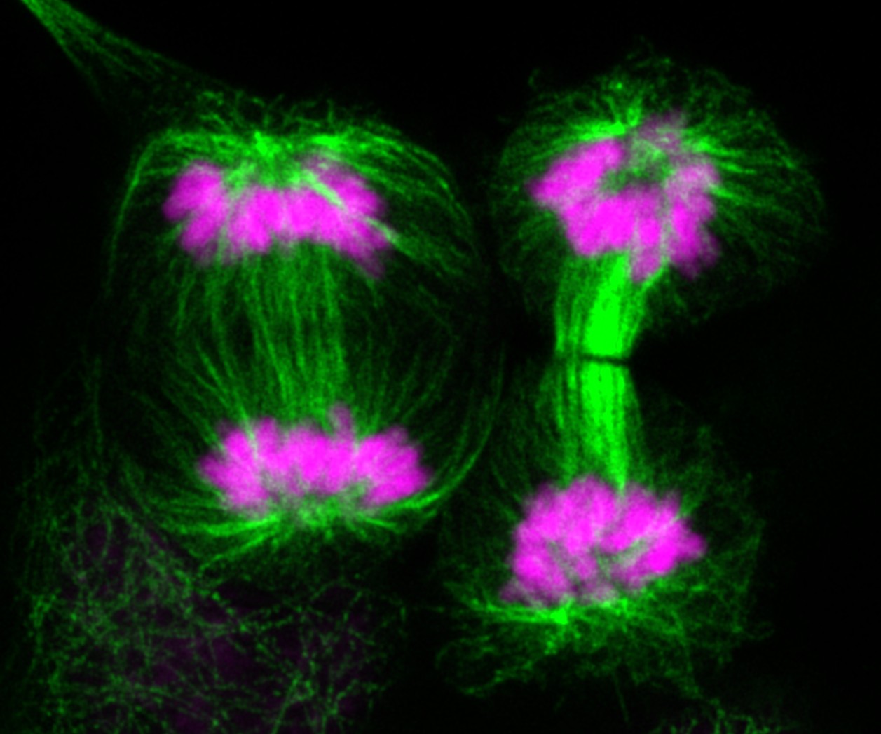
Mirror mirror on the wall, how beautiful is cell division after all?

Image: Tubulin (green) and DAPI (magenta) staining in H2228 lung cancer cells. This multiple image projection was acquired using Zeiss LSM 980 Airyscan confocal laser scanning microscope, and 63x objective, and processed using lmageJ software.
This image shows two cancer cells dividing happily. These symmetrical events might look nice in the picture, but it is not what we want to happen in cancer patients' tumours. Maria and her colleagues are working on drugs targeting regulators of cell division (directly or indirectly), which might provide an effective way to stop cancer in its tracks.
Julian Hughes, former Chief Photographer at Trinity Mirror Group plc and one of the judges, said of the winning image:
“This captivating image, with its striking symmetry and vivid contrast, offers both aesthetic beauty and scientific depth.
The dual staining of tubulin and DNA in H2228 lung cancer cells is not only technically impressive but also emotionally resonant – a visual paradox of elegance and pathology. The image demonstrates a high level of technical skill – the clarity and resolution achieved at 63x magnification allow viewers to appreciate the intricacies of mitosis in cancer cells.
However, what truly set this entry apart was its narrative power. The title cleverly juxtaposes fairy tale imagery with the harsh reality of cancer biology, inviting reflection on the beauty and danger of unchecked cell division. It’s a compelling reminder that scientific images can be both informative and poetic.”
Second place
Dr Mateus Crespo, Dr Bora Gurel and Professor Johann de Bono in the Cancer Biomarkers Group in the Division of Clinical Studies.
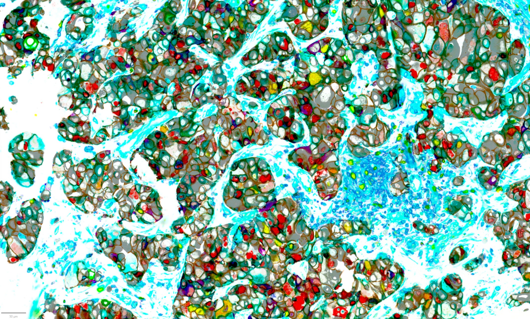
The Shape of Resistance

Image: An analysis of 40 biomarkers in a single advanced prostate cancer tumour sample showing how the different cell types are organised and interconnected.
The researchers have spent many years studying the complex makeup of metastatic, castration-resistant prostate cancer (mCRPC). The focus is not solely on the cancer itself, but also on the surrounding microenvironment — including how different types of cells are arranged and how the immune system is involved. The aim is to better understand tumour heterogeneity (the differences within and between tumours), lineage plasticity (how cancer cells can change their identity), and the immune microenvironment in mCRPC.
To capture this complexity, advanced tools have been developed that simultaneously analyse over 40 biomarkers in a single tumour sample. Using a technique known as hyperplex immunofluorescence, we can now visualise how various cell types are organised and interconnected within the tumour's ecosystem.
This technology offers valuable insights into how advanced prostate tumours evolve and adapt under treatment pressure.
Third place
Dr Amin Mirza, Head of the Structural Chemistry Facility in the Centre for Cancer Drug Discovery.
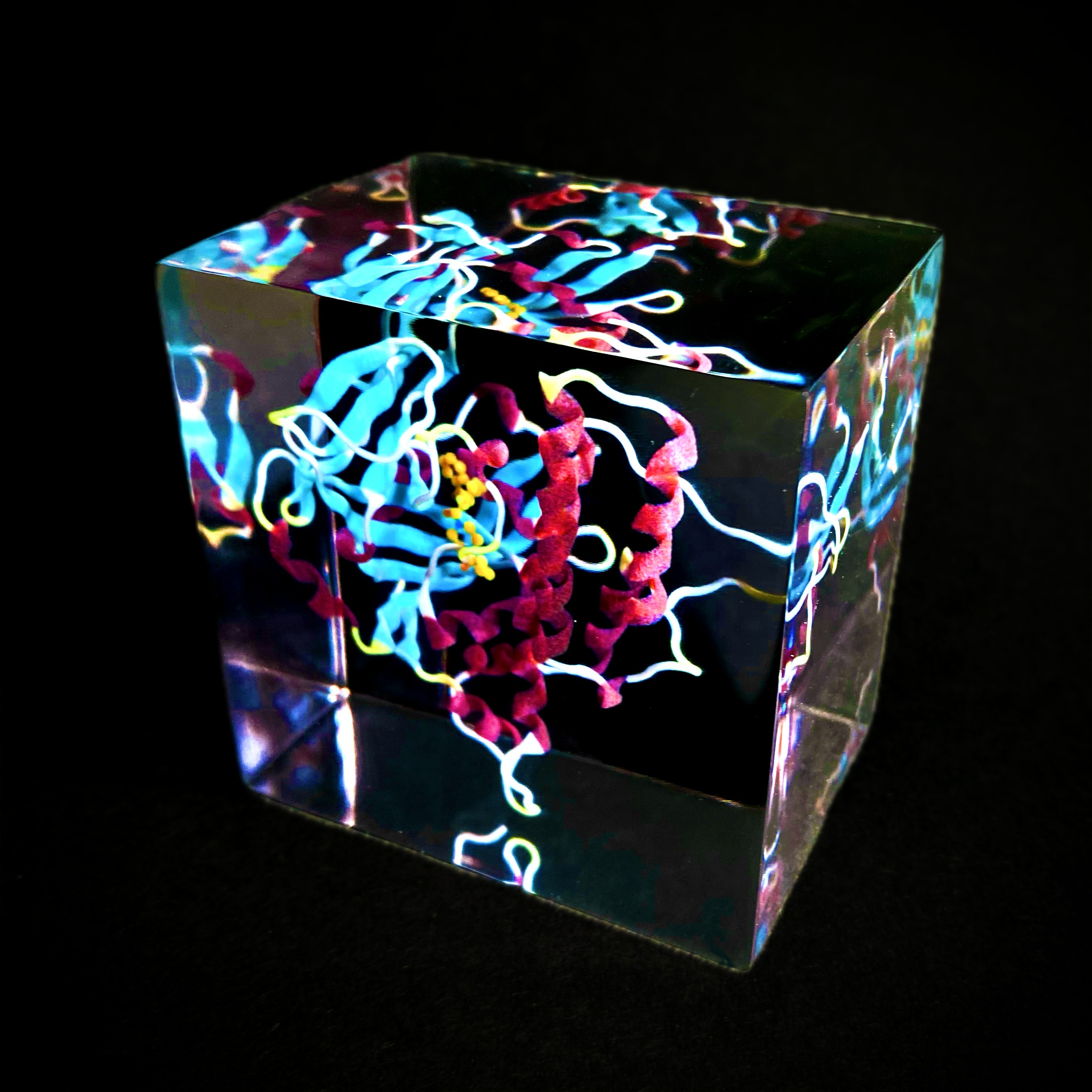
My claret and blue heart

Image: 3D-printed model of the targeted PARP-inhibitor drug Olaparib.
This is a 3D-printed model of the targeted drug olaparib (yellow) within the protein PARP1 which is depicted using ribbons and helices in the West Ham United FC colours. It was printed by Craig Cummings on a MediJet_J5 3D printer.
The image shows how the drug binds at the ‘heart’ of the PARP1 protein and by coincidence the protein resembles a heart from various angles. Olaparib is used in the treatment of ovarian, breast, pancreatic and prostate cancers. It works by blocking the PARP protein which leads to cancer cell death.
Researchers at the ICR played a key role in the development of PARP inhibitors , including olaparib.
Social media winner
Dr Qiyun Zhong, Postdoctoral Training Fellow in the Dynamical Cell Systems Group in the Division of Cell and Molecular Biology.
.png?sfvrsn=449853ce_5)
Image: E. coli bacteria (blue dots) engineered with microscopic ‘stings’ (red) to deliver treatment to pancreatic cancer cells (green). It was imaged by a Carl Zeiss Z1 microscope at 100x.
This image shows engineered E. coli bacteria (blue dots) that are decorated with microscopic ‘stings’ (red) to deliver a cocktail of ‘poisons’ to pancreatic cancer cells. The bacteria completely take over the pancreatic cancer cellular structure (green) forcing cancer cells to distort, shrink and die, and at the same time release signals to boost the immune system.
Pancreatic cancer is one of the deadliest cancer types and very limited treatments exist. The efficiency of these engineered bacteria to eliminate pancreatic cancer cells and trigger beneficial immune responses is much superior than even high doses of available cancer therapy agents, and they could inspire the next generation of cancer therapy development.
This entry was chosen from the shortlist shared on the ICR’s social media channels and received half of the total votes.
Judging panel
The judging panel for the ICR Science and Medical Image Competition was made up of:
- Professor Udai Banerji, Co-Director of the Drug Development Unit: Clinical Pharmacodynamics Biomarker Group, Clinical Pharmacology & Trials, Clinical Pharmacology Adaptive Therapy Goup
- Dr Benjamin Hartley, Arts Officer at The Royal Marsden
- Eleanor Howard, Head of Strategic Marketing at the ICR
- Dr Tina Daviter, Head of Core Research Facilities
- Julian Hughes, formerly Chief Photographer at Trinity Mirror Group plc before his retirement.
Cutting-edge research
These eye-catching images illustrate just some of the cutting-edge research carried out at the ICR.
From images like these our researchers are gaining unprecedented insights into the mechanisms that drive cancer, to then discover new ways to target the disease and help treat patients.
Help the ICR’s researchers continue to make the discoveries that defeat cancer.
Donate now