At The Institute of Cancer Research, London, our ability to visualise the intricate inner workings of cancer is going from strength to strength. Robbie Lockyer spoke with scientists using cutting-edge imaging techniques to uncover how these tools are helping us understand cancer in unprecedented detail.
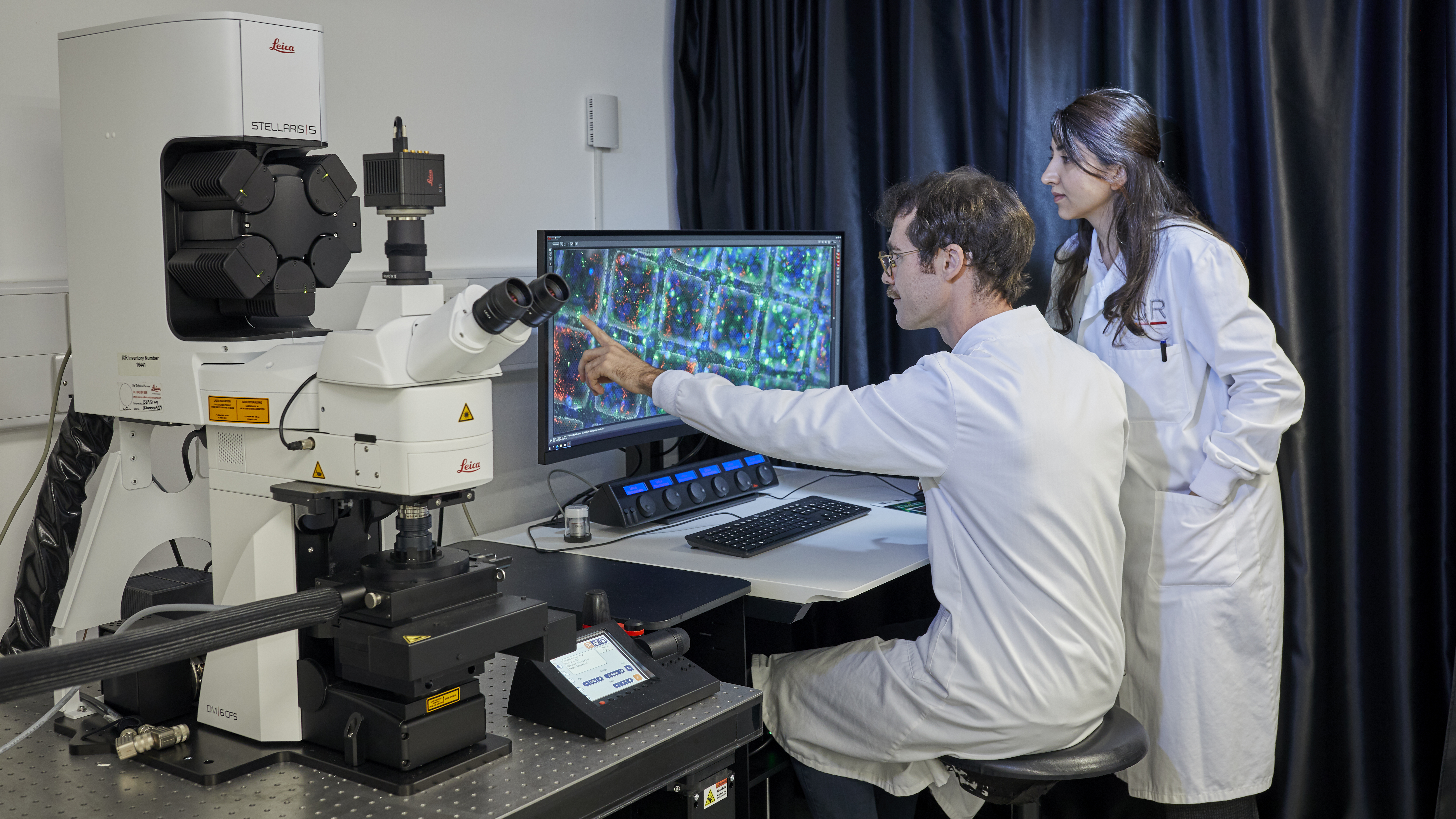
From high-throughput robotics to cryogenic electron microscopes, the state-of-the-art imaging technologies housed at The Institute of Cancer Research (ICR) are world-class. One recent addition – a confocal microscope called the STELLARIS – is helping researchers visualise life at the molecular level inside cancer cells, providing a critical bridge between cell biology and structural biology.
It’s one of the most sophisticated pieces of imaging technology in the ICR’s collection – and it’s helping us understand how cancer really works.
Why is this needed?
If you imagine cancer as a disease of misbehaving proteins, you quickly realise how important it is to see where those proteins are, how they interact and what happens when they malfunction. Until recently, most structural biologists have studied proteins in isolation, meaning they are highly purified in test tubes.
Dr Matthew Jessop, Senior Scientific Officer at the ICR, said: “Proteins don’t exist alone in the body. They operate in the crowded, complex environment of the cell. To understand how they behave, and misbehave, we need to see them where they naturally live.”
That’s where the STELLARIS comes in. It uses a tiny pinhole to illuminate a single section within the cell, rather than lighting up the whole sample at once. Researchers can focus on a thin slice, which allows them to very precisely locate proteins within a cell. This kind of visualisation is vital for understanding complex biological systems, especially in cancer where protein mislocalisation or malfunction can play a key role in disease progression.
Building a powerful imaging pipeline
The STELLARIS microscope facilitates the first step in a cutting-edge process known as Correlated Light and Electron Microscopy (CLEM), which is now supported by a dedicated CLEM Lab at the ICR. This process allows researchers to use light microscopy – used for tracking fluorescently labelled proteins to locate specific proteins and then study them in ultra-fine detail using electron microscopy, which allows researchers to build up highly detailed, three-dimensional reconstructions of the structures inside cells.
Dr Teige Matthews-Palmer, Electron Microscopy Facility Manager in the Structural Biology Division at the ICR, said: “We tag the protein we’re interested in with a fluorescent marker. The STELLARIS shows us exactly where that protein is within the cell. Then, we take the sample to the electron microscope and zoom in on that exact spot. It’s like using a map to find treasure.”
Once the target is located, the sample can be frozen, sectioned and examined at atomic or near-atomic resolution using cryo-electron microscopy, giving us a view into the real-life context of these proteins.
Dr Matthews-Palmer said: “When you look at an entire cell in the electron microscope, it’s packed full of things. If we’re looking for a particular protein of interest, it’s like trying to find a needle in a haystack.”
With confocal imaging as the guide, researchers combine the specificity of fluorescence with the resolution of electron microscopy to create a hybrid workflow that gives them the best of both worlds.
The CLEM lab
The CLEM lab was established with funding from the Wolfson Foundation as part of a broader investment into equipment that helps structural biologists explore proteins inside cells – not just in purified tubes.
The lab is jointly supported by the light and electron microscopy facilities. Dr Jessop operates the STELLARIS while Dr Matthews-Palmer oversees the broader electron microscopy setup. Together, they’re building a facility that bridges expertise across disciplines.
Dr Matthews-Palmer said: “The use of confocal microscopy isn’t restricted to a single study of cancer. It’s particularly used by structural biologists, but it represents a bridge between structural and cell biology. There’s both an opportunity, and need, for more collaboration between these fields at the ICR.”
One example comes from the Structural Biology of Cell Signalling Group, who are investigating a protein called tankyrase, which is known to play a role in cancer development. Using the STELLARIS microscope, researchers are able to visualise tagged tankyrase molecules in living cells, rather than just in purified test tubes, helping them select precise regions for ultra-thin sectioning and electron imaging for analysis.
Dr Jessop said: “We freeze the cells to vitrify them – turning the water in cells to glass without forming ice crystals. Then we use a focused ion beam to carve out a window just nanometres thick, allowing us to see inside the cell with incredible resolution.”
A powerful tool
The STELLARIS isn’t a simple plug-and-play machine. It requires careful handling, expert training and delicate sample preparation. The samples must be frozen, stored at -196°C and handled on delicate wafer-thin grids. They also need to be protected from moisture in the air, which can destroy a sample.
Because of this, researchers need to consult with the lab’s technical specialists before starting. For long-term use, researchers can undergo training to become independent users.
Dr Matthews-Palmer said: “We want to try to get as many researchers using the equipment as we can. We’re currently looking for projects to test it out further and see what we can do. It’s a big deal for the ICR to have it.
“Before this, you could only study purified proteins outside the cell. Now, we can track those same proteins inside tissues or organoids – see how they behave, where they move and what other molecules they interact with.”
Looking ahead: a new microscope for an even higher level of detail
The team hopes to build on the STELLARIS’s capabilities with a focused ion beam scanning electron microscope (FIB-SEM) – a £1.5 million piece of equipment that, unlike the STELLARIS which uses light to locate proteins, would allow researchers to slice and scan samples in three dimensions with nanometre precision.
Imagine trying to shine a torch through your hand – nothing gets through. But if you shine it through your fingertip, you see a red glow. That’s the idea behind the new system, it cuts wafer-thin slices, or windows, so that electrons can pass through and reveal molecular detail anywhere in the cell.
Dr Matthews-Palmer said: “We’ve just been awarded funding from the Biotechnology and Biological Sciences Research Council to obtain the new FIB-SEM at the ICR, which we will make available to other researchers in London. To have the ability to carry out the full workflow of structural biology inside cells will be very exciting and push our research further forward."
Dr Jessop said: “It’s a cutting-edge technique that means we can see exactly where the atoms are in a molecule, deep inside the cell.
“This microscope would complete the imaging pipeline that would position the ICR at the forefront of cellular structural biology – not just in the UK, but globally.”
Powered by support
Advanced technologies like the STELLARIS – and the groundbreaking research it enables – are made possible by the generosity of donors, foundations and supporters who share our mission to defeat cancer. While the platform is still in its early stages, staff are already seeing exciting potential.
With continued investment, we can expand this powerful imaging pipeline and accelerate our understanding of cancer from the inside out – one molecule at a time.
With your generous support, we can continue making more discoveries, finding more cures, and saving more lives.
Make a donation today