Paediatric Solid Tumour Biology and Therapeutics Group

Professor Louis Chesler’s group is investigating the genetic causes for the childhood cancers, neuroblastoma, medulloblastoma and rhabdomyosarcoma. 

Research, projects and publications in this group

Our group's aim is to improve the treatment and survival of children with neuroblastoma, medulloblastoma and rhabdomyosarcoma.

The goal of our laboratory is to improve the treatment and survival of children with neuroblastoma, medulloblastoma and rhabdomyosarcoma, three paediatric solid tumours in which high-risk patient cohorts can be defined by alterations in a single oncogene. We focus on the role of the MYCN oncogene, since aberrant expression of MYCNis very significantly associated with high-risk in all three diseases and implies that they may have a common cell-of-origin.

Elucidating the molecular signalling pathways that control expression of the MYCN oncoprotein and targeting these pathways with novel therapeutics is a major goal of the laboratory. We use a variety of innovative preclinical drug development platforms for this purpose.

Technologically, we focus on genetically engineered cancer models incorporating novel imaging (optical and fluorescent) modalities that can be used as markers to monitor disease progression and therapeutic response.

Our group has several key objectives:

  • Mechanistically dissect the role of the MYCN oncogene, and other key oncogenic driver genes in poor-outcome paediatric solid tumours (neuroblastoma, medulloblastoma, rhabdomyosarcoma).
  • Develop novel therapeutics targeting MYCN oncoproteins and other key oncogenic drivers
  • Develop improved genetic cancer models dually useful for studies of oncogenesis and preclinical development of novel therapeutics.
  • Use such models to develop and functionally validate optical imaging modalities useful as surrogate markers of tumour progression in paediatric cancer.

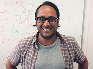
Professor Louis Chesler

Clinical Senior Lecturer/Group Leader:

Paediatric Solid Tumour Biology and Therapeutics Professor Louis Chesler (Profile pic)

Professor Louis Chesler is working to understand the biology of children’s cancers and use that information to discover and develop new personalised approaches to cancer treatment. His work focuses on improving the understanding of the role of the MYCN oncogene.

Researchers in this group

.

Email: [email protected]

Location: Sutton

.

Phone: +44 20 3437 6124

Email: [email protected]

Location: Sutton

.

Email: [email protected]

Location: Sutton

.

Email: [email protected]

Location: Sutton

.

Phone: +44 20 3437 3617

Email: [email protected]

Location: Sutton

.

Phone: +44 20 8722 4186

Email: [email protected]

Location: Sutton

.

Phone: +44 20 3437 3501

Email: [email protected]

Location: Sutton

.

Email: [email protected]

Location: Sutton

.

Phone: +44 20 8722 4361

Email: [email protected]

Location: Sutton

.

Email: [email protected]

Location: Sutton

.

Phone: +44 20 3437 6118

Email: [email protected]

Location: Sutton

.

Phone: +44 20 3437 6021

Email: [email protected]

Location: Sutton

.

Phone: +44 20 3437 6196

Email: [email protected]

Location: Sutton

.

Phone: +44 20 3437 6258

Email: [email protected]

Location: Sutton

.

Email: [email protected]

Location: Sutton

.

Phone: +44 20 3437 6121

Email: [email protected]

Location: Sutton

.

Phone: +44 20 8722 4527

Email: [email protected]

Location: Sutton

.

OrcID: 0000-0003-3977-7020

Phone: +44 20 3437 6109

Email: [email protected]

Location: Sutton

I obtained an MSci in Biochemistry from the University of Glasgow in 2018. In October 2018 I joined the labs of Dr Michael Hubank and Professor Andrea Sottoriva to investigate the use of liquid biopsy to monitor clonal frequency and emergence of resistance mutations in paediatric cancers.

.

Email: [email protected]

Location: Sutton

.

Phone: +44 20 3437 6358

Email: [email protected]

Location: Sutton

.

Phone: +44 20 3437 6131

Email: [email protected]

Location: Sutton

.

Email: [email protected]

Location: Sutton

Professor Louis Chesler's group have written 113 publications

Most recent new publication 4/2025

See all their publications

Vacancies in this group

Working in this group

Prospect Research Manager

  • Chelsea
  • Development and Communications Directorate
  • £41,600 - £43,000
  • Permanent

About the Role Our Prospect Research team plays a central role in unlocking new funding opportunities across the ICR's philanthropic income streams. It is responsible for delivering high quality, insightful prospect briefings and fundraising intelligence, which supports the ICR's fundraisers in their efforts to build meaningful relationships with philanthropic high net-worth individuals, trusts, foundations and corporates who have the interest and capacity to support the ICR's vital research. The post holder will play a key role in supporting the Development & Communications directorate to source, assess and validate new funding opportunities, maximise income secured across our philanthropic income streams and fundraising campaign, and grow our incredible donor community. The successful candidate will have experience of devising and successfully implementing targeted prospecting strategies. You will play a lead role in identifying philanthropic HNWIs and corporates with the capacity and inclination to support our work. The post holder will also be responsible for carrying out due diligence research and alerting fundraisers to reputational risks identified in support of the ICR's gift acceptance policy. You will work closely with the Head of Prospect Development to support fundraisers in maximising the success of our senior volunteer network through network mapping and expanding its ranks. In addition, the post holder will manage the Prospect Research Executive, providing development and mentoring support. What We Offer A supportive and collaborative working environment. Opportunities for professional development and career progression. Competitive salary and pension We encourage all applicants to access the job pack attached for more detailed information regarding this role. For an informal discussion regarding the role, please contact Caroline Porter via telephone on 020 7153 5486 or email at [email protected].

Principal Statistician

  • Sutton
  • Clinical Trials and Statistics Unit
  • £49,970 - £63,120 per annum
  • Fixed term

Job Details The Cancer Research UK funded Clinical Trials and Statistics Unit at The Institute of Cancer Research (ICR-CTSU) seek an experienced and highly motivated Principal Statistician to join their team of over 20 statisticians. We want to hear from you if you: Enjoy the challenge of designing efficient clinical trials and analysing complex datasets to answer clinical questions that will make a difference in patients’ lives Seek variety in your work and opportunities to apply your statistical knowledge across multiple therapeutic areas in oncology. Thrive on being part of a multi-disciplinary research team with like-minded statisticians. Are looking to develop your career within a dynamic and supportive academic environment at a leading cancer clinical trials unit. Key Requirements The successful applicant will be an experienced and highly motivated medical statistician interested in applying their statistical knowledge to the design and analysis of patient-centred clinical trials. They will have a post-graduate qualification in statistics. They should demonstrate a solid understanding of clinical trials and experience in applying statistical methods to real-world data. Effective oral and written communication skills, as well as enthusiasm for collaborating with others from diverse disciplines, are essential. Department/Directorate Information ICR-CTSU manages an exciting portfolio of national and international phase II and III cancer clinical trials and an expanding number of phase I trials. You will work as part of a multi-disciplinary team on the statistical design and development of new trials. You will conduct and oversee analysis of a number of clinical trials and/ or associated imaging or translational biomarker research studies across our portfolio. In your supporting statement please summarise how your research/managerial experience fits with the role. We encourage all applicants to access the job pack attached for more detailed information regarding this role. For an informal discussion about the role please contact Nuria Porta, email [email protected] This is an office based role. Requests for hybrid working (splitting time between our Sutton site and home) may be considered following successful completion of key training and only if the role allows. Flexible working options may be considered. Informal enquiries may be made by emailing [email protected]

Industrial partnership opportunities with this group

Opportunity: A novel test for predicting future cancer risk in patients with inflammatory bowel disease

Commissioner: Professor Trevor Graham

Recent discoveries from this group

21/08/25

Seven images were shortlisted for this year’s annual Science and Medical Image competition, showcasing the eye-catching science being carried out at the ICR. Three winners were selected by a judging panel and the fourth was chosen by the public based on votes on social media.

Images of cancer research

Our annual imaging competition celebrates the incredible research by our scientists at the ICR and The Royal Marsden, and the arresting images they produce – often they are capturing cellular processes in action, which is key to making new discoveries or deepening our understanding of clinical problems affecting cancer patients.

The selection chosen for the judges’ shortlist covers different cancer types and different scientific techniques such as immunofluorescence, microscopy and 3D printing.

Judges’ winner

Dr Maria Taskinen, Postdoctoral Training Fellow in the Signal Transduction and Molecular Pharmacology Group in the Division of Cancer Therapeutics.

Mirror mirror on the wall, how beautiful is cell division after all?
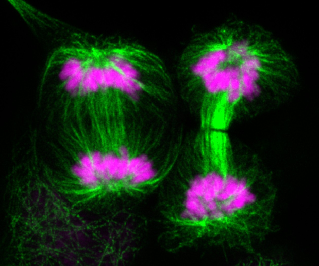
Tubulin (green) and DAPI (magenta) staining in H2228 lung cancer cells

Image: Tubulin (green) and DAPI (magenta) staining in H2228 lung cancer cells. This multiple image projection was acquired using Zeiss LSM 980 Airyscan confocal laser scanning microscope, and 63x objective, and processed using lmageJ software.

This image shows two cancer cells dividing happily. These symmetrical events might look nice in the picture, but it is not what we want to happen in cancer patients' tumours. Maria and her colleagues are working on drugs targeting regulators of cell division (directly or indirectly), which might provide an effective way to stop cancer in its tracks.

Julian Hughes, former Chief Photographer at Trinity Mirror Group plc and one of the judges, said of the winning image:

“This captivating image, with its striking symmetry and vivid contrast, offers both aesthetic beauty and scientific depth.

The dual staining of tubulin and DNA in H2228 lung cancer cells is not only technically impressive but also emotionally resonant – a visual paradox of elegance and pathology. The image demonstrates a high level of technical skill – the clarity and resolution achieved at 63x magnification allow viewers to appreciate the intricacies of mitosis in cancer cells.

However, what truly set this entry apart was its narrative power. The title cleverly juxtaposes fairy tale imagery with the harsh reality of cancer biology, inviting reflection on the beauty and danger of unchecked cell division. It’s a compelling reminder that scientific images can be both informative and poetic.”

Second place

Dr Mateus Crespo, Dr Bora Gurel and Professor Johann de Bono in the Cancer Biomarkers Group in the Division of Clinical Studies.

The Shape of Resistance
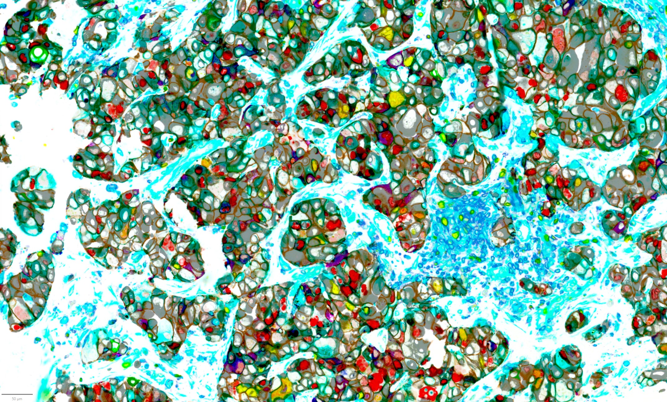
An analysis of 40 biomarkers in a single advanced prostate cancer tumour sample showing how the different cell types are organised and interconnected

Image: An analysis of 40 biomarkers in a single advanced prostate cancer tumour sample showing how the different cell types are organised and interconnected.

The researchers have spent many years studying the complex makeup of metastatic, castration-resistant prostate cancer (mCRPC). The focus is not solely on the cancer itself, but also on the surrounding microenvironment — including how different types of cells are arranged and how the immune system is involved. The aim is to better understand tumour heterogeneity (the differences within and between tumours), lineage plasticity (how cancer cells can change their identity), and the immune microenvironment in mCRPC.

To capture this complexity, advanced tools have been developed that simultaneously analyse over 40 biomarkers in a single tumour sample. Using a technique known as hyperplex immunofluorescence, we can now visualise how various cell types are organised and interconnected within the tumour's ecosystem.

This technology offers valuable insights into how advanced prostate tumours evolve and adapt under treatment pressure.

Third place

Dr Amin Mirza, Head of the Structural Chemistry Facility in the Centre for Cancer Drug Discovery. 

My claret and blue heart
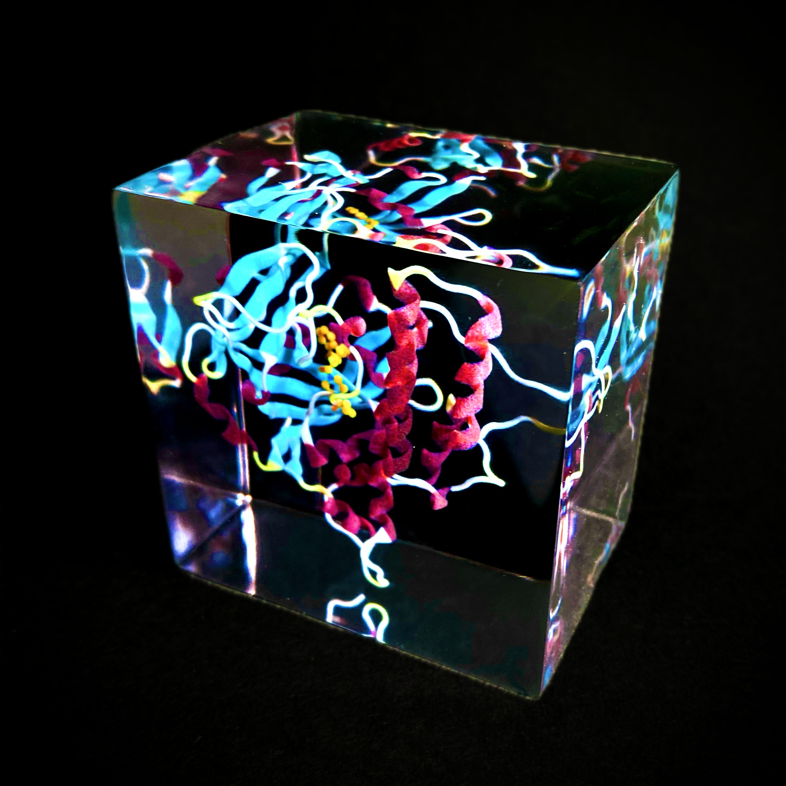
3D-printed model of the targeted PARP-inhibitor drug Olaparib

Image: 3D-printed model of the targeted PARP-inhibitor drug Olaparib.

This is a 3D-printed model of the targeted drug olaparib (yellow) within the protein PARP1 which is depicted using ribbons and helices in the West Ham United FC colours. It was printed by Craig Cummings on a MediJet_J5 3D printer. 

The image shows how the drug binds at the ‘heart’ of the PARP1 protein and by coincidence the protein resembles a heart from various angles. Olaparib is used in the treatment of ovarian, breast, pancreatic and prostate cancers. It works by blocking the PARP protein which leads to cancer cell death.

Researchers at the ICR played a key role in the development of PARP inhibitors , including olaparib.

Social media winner

Dr Qiyun Zhong, Postdoctoral Training Fellow in the Dynamical Cell Systems Group in the Division of Cell and Molecular Biology.

E. coli bacteria engineered with microscopic ‘stings’ to deliver treatment to pancreatic cancer cells

Image: E. coli bacteria (blue dots) engineered with microscopic ‘stings’ (red) to deliver treatment to pancreatic cancer cells (green). It was imaged by a Carl Zeiss Z1 microscope at 100x.

This image shows engineered E. coli bacteria (blue dots) that are decorated with microscopic ‘stings’ (red) to deliver a cocktail of ‘poisons’ to pancreatic cancer cells. The bacteria completely take over the pancreatic cancer cellular structure (green) forcing cancer cells to distort, shrink and die, and at the same time release signals to boost the immune system. 

Pancreatic cancer is one of the deadliest cancer types and very limited treatments exist. The efficiency of these engineered bacteria to eliminate pancreatic cancer cells and trigger beneficial immune responses is much superior than even high doses of available cancer therapy agents, and they could inspire the next generation of cancer therapy development.

This entry was chosen from the shortlist shared on the ICR’s social media channels and received half of the total votes.

Judging panel

The judging panel for the ICR Science and Medical Image Competition was made up of: 

  • Professor Udai Banerji, Co-Director of the Drug Development Unit: Clinical Pharmacodynamics Biomarker Group, Clinical Pharmacology & Trials, Clinical Pharmacology Adaptive Therapy Goup
  • Dr Benjamin Hartley, Arts Officer at The Royal Marsden
  • Eleanor Howard, Head of Strategic Marketing at the ICR 
  • Dr Tina Daviter, Head of Core Research Facilities 
  • Julian Hughes, formerly Chief Photographer at Trinity Mirror Group plc before his retirement. 

Cutting-edge research

These eye-catching images illustrate just some of the cutting-edge research carried out at the ICR.

From images like these our researchers are gaining unprecedented insights into the mechanisms that drive cancer, to then discover new ways to target the disease and help treat patients.

Help the ICR’s researchers continue to make the discoveries that defeat cancer.

Donate now