Paediatric Solid Tumour Biology and Therapeutics Group

Professor Louis Chesler’s group is investigating the genetic causes for the childhood cancers, neuroblastoma, medulloblastoma and rhabdomyosarcoma. 

Research, projects and publications in this group

Our group's aim is to improve the treatment and survival of children with neuroblastoma, medulloblastoma and rhabdomyosarcoma.

The goal of our laboratory is to improve the treatment and survival of children with neuroblastoma, medulloblastoma and rhabdomyosarcoma, three paediatric solid tumours in which high-risk patient cohorts can be defined by alterations in a single oncogene. We focus on the role of the MYCN oncogene, since aberrant expression of MYCNis very significantly associated with high-risk in all three diseases and implies that they may have a common cell-of-origin.

Elucidating the molecular signalling pathways that control expression of the MYCN oncoprotein and targeting these pathways with novel therapeutics is a major goal of the laboratory. We use a variety of innovative preclinical drug development platforms for this purpose.

Technologically, we focus on genetically engineered cancer models incorporating novel imaging (optical and fluorescent) modalities that can be used as markers to monitor disease progression and therapeutic response.

Our group has several key objectives:

  • Mechanistically dissect the role of the MYCN oncogene, and other key oncogenic driver genes in poor-outcome paediatric solid tumours (neuroblastoma, medulloblastoma, rhabdomyosarcoma).
  • Develop novel therapeutics targeting MYCN oncoproteins and other key oncogenic drivers
  • Develop improved genetic cancer models dually useful for studies of oncogenesis and preclinical development of novel therapeutics.
  • Use such models to develop and functionally validate optical imaging modalities useful as surrogate markers of tumour progression in paediatric cancer.

Professor Louis Chesler

Clinical Senior Lecturer/Group Leader:

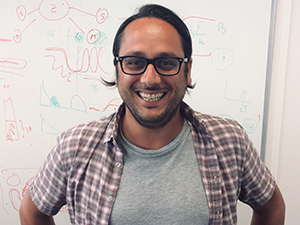
Paediatric Solid Tumour Biology and Therapeutics Professor Louis Chesler (Profile pic)

Professor Louis Chesler is working to understand the biology of children’s cancers and use that information to discover and develop new personalised approaches to cancer treatment. His work focuses on improving the understanding of the role of the MYCN oncogene.

Researchers in this group

.

Email: [email protected]

Location: Sutton

.

Phone: +44 20 3437 6124

Email: [email protected]

Location: Sutton

.

Email: [email protected]

Location: Sutton

.

Email: [email protected]

Location: Sutton

.

Phone: +44 20 3437 3617

Email: [email protected]

Location: Sutton

.

Phone: +44 20 8722 4186

Email: [email protected]

Location: Sutton

.

Phone: +44 20 3437 3501

Email: [email protected]

Location: Sutton

.

Email: [email protected]

Location: Sutton

.

Phone: +44 20 8722 4361

Email: [email protected]

Location: Sutton

.

Email: [email protected]

Location: Sutton

.

Phone: +44 20 3437 6118

Email: [email protected]

Location: Sutton

.

Phone: +44 20 3437 6021

Email: [email protected]

Location: Sutton

.

Phone: +44 20 3437 6196

Email: [email protected]

Location: Sutton

.

Phone: +44 20 3437 6258

Email: [email protected]

Location: Sutton

.

Email: [email protected]

Location: Sutton

.

Phone: +44 20 3437 6121

Email: [email protected]

Location: Sutton

.

Phone: +44 20 8722 4527

Email: [email protected]

Location: Sutton

.

OrcID: 0000-0003-3977-7020

Phone: +44 20 3437 6109

Email: [email protected]

Location: Sutton

I obtained an MSci in Biochemistry from the University of Glasgow in 2018. In October 2018 I joined the labs of Dr Michael Hubank and Professor Andrea Sottoriva to investigate the use of liquid biopsy to monitor clonal frequency and emergence of resistance mutations in paediatric cancers.

.

Email: [email protected]

Location: Sutton

.

Phone: +44 20 3437 6358

Email: [email protected]

Location: Sutton

.

Phone: +44 20 3437 6131

Email: [email protected]

Location: Sutton

.

Email: [email protected]

Location: Sutton

Professor Louis Chesler's group have written 113 publications

Most recent new publication 4/2025

See all their publications

Vacancies in this group

Working in this group

Prospect Research Manager

  • Chelsea
  • Development and Communications Directorate
  • £41,600 - £43,000
  • Permanent

About the Role Our Prospect Research team plays a central role in unlocking new funding opportunities across the ICR's philanthropic income streams. It is responsible for delivering high quality, insightful prospect briefings and fundraising intelligence, which supports the ICR's fundraisers in their efforts to build meaningful relationships with philanthropic high net-worth individuals, trusts, foundations and corporates who have the interest and capacity to support the ICR's vital research. The post holder will play a key role in supporting the Development & Communications directorate to source, assess and validate new funding opportunities, maximise income secured across our philanthropic income streams and fundraising campaign, and grow our incredible donor community. The successful candidate will have experience of devising and successfully implementing targeted prospecting strategies. You will play a lead role in identifying philanthropic HNWIs and corporates with the capacity and inclination to support our work. The post holder will also be responsible for carrying out due diligence research and alerting fundraisers to reputational risks identified in support of the ICR's gift acceptance policy. You will work closely with the Head of Prospect Development to support fundraisers in maximising the success of our senior volunteer network through network mapping and expanding its ranks. In addition, the post holder will manage the Prospect Research Executive, providing development and mentoring support. What We Offer A supportive and collaborative working environment. Opportunities for professional development and career progression. Competitive salary and pension We encourage all applicants to access the job pack attached for more detailed information regarding this role. For an informal discussion regarding the role, please contact Caroline Porter via telephone on 020 7153 5486 or email at [email protected].

Principal Statistician

  • Sutton
  • Clinical Trials and Statistics Unit
  • £49,970 - £63,120 per annum
  • Fixed term

Job Details The Cancer Research UK funded Clinical Trials and Statistics Unit at The Institute of Cancer Research (ICR-CTSU) seek an experienced and highly motivated Principal Statistician to join their team of over 20 statisticians. We want to hear from you if you: Enjoy the challenge of designing efficient clinical trials and analysing complex datasets to answer clinical questions that will make a difference in patients’ lives Seek variety in your work and opportunities to apply your statistical knowledge across multiple therapeutic areas in oncology. Thrive on being part of a multi-disciplinary research team with like-minded statisticians. Are looking to develop your career within a dynamic and supportive academic environment at a leading cancer clinical trials unit. Key Requirements The successful applicant will be an experienced and highly motivated medical statistician interested in applying their statistical knowledge to the design and analysis of patient-centred clinical trials. They will have a post-graduate qualification in statistics. They should demonstrate a solid understanding of clinical trials and experience in applying statistical methods to real-world data. Effective oral and written communication skills, as well as enthusiasm for collaborating with others from diverse disciplines, are essential. Department/Directorate Information ICR-CTSU manages an exciting portfolio of national and international phase II and III cancer clinical trials and an expanding number of phase I trials. You will work as part of a multi-disciplinary team on the statistical design and development of new trials. You will conduct and oversee analysis of a number of clinical trials and/ or associated imaging or translational biomarker research studies across our portfolio. In your supporting statement please summarise how your research/managerial experience fits with the role. We encourage all applicants to access the job pack attached for more detailed information regarding this role. For an informal discussion about the role please contact Nuria Porta, email [email protected] This is an office based role. Requests for hybrid working (splitting time between our Sutton site and home) may be considered following successful completion of key training and only if the role allows. Flexible working options may be considered. Informal enquiries may be made by emailing [email protected]

Industrial partnership opportunities with this group

Opportunity: A novel test for predicting future cancer risk in patients with inflammatory bowel disease

Commissioner: Professor Trevor Graham

Recent discoveries from this group

07/08/25 - by

At The Institute of Cancer Research, London, our ability to visualise the intricate inner workings of cancer is going from strength to strength. Robbie Lockyer spoke with scientists using cutting-edge imaging techniques to uncover how these tools are helping us understand cancer in unprecedented detail.

From high-throughput robotics to cryogenic electron microscopes, the state-of-the-art imaging technologies housed at The Institute of Cancer Research (ICR) are world-class. One recent addition – a confocal microscope called the STELLARIS – is helping researchers visualise life at the molecular level inside cancer cells, providing a critical bridge between cell biology and structural biology.

It’s one of the most sophisticated pieces of imaging technology in the ICR’s collection – and it’s helping us understand how cancer really works.

Why is this needed?

If you imagine cancer as a disease of misbehaving proteins, you quickly realise how important it is to see where those proteins are, how they interact and what happens when they malfunction. Until recently, most structural biologists have studied proteins in isolation, meaning they are highly purified in test tubes.

Dr Matthew Jessop, Senior Scientific Officer at the ICR, said: “Proteins don’t exist alone in the body. They operate in the crowded, complex environment of the cell. To understand how they behave, and misbehave, we need to see them where they naturally live.”

That’s where the STELLARIS comes in. It uses a tiny pinhole to illuminate a single section within the cell, rather than lighting up the whole sample at once. Researchers can focus on a thin slice, which allows them to very precisely locate proteins within a cell. This kind of visualisation is vital for understanding complex biological systems, especially in cancer where protein mislocalisation or malfunction can play a key role in disease progression.  

Building a powerful imaging pipeline

The STELLARIS microscope facilitates the first step in a cutting-edge process known as Correlated Light and Electron Microscopy (CLEM), which is now supported by a dedicated CLEM Lab at the ICR. This process allows researchers to use light microscopy – used for tracking fluorescently labelled proteins to locate specific proteins and then study them in ultra-fine detail using electron microscopy, which allows researchers to build up highly detailed, three-dimensional reconstructions of the structures inside cells.

Dr Teige Matthews-Palmer, Electron Microscopy Facility Manager in the Structural Biology Division at the ICR, said: “We tag the protein we’re interested in with a fluorescent marker. The STELLARIS shows us exactly where that protein is within the cell. Then, we take the sample to the electron microscope and zoom in on that exact spot. It’s like using a map to find treasure.”

Once the target is located, the sample can be frozen, sectioned and examined at atomic or near-atomic resolution using cryo-electron microscopy, giving us a view into the real-life context of these proteins.

Dr Matthews-Palmer said: “When you look at an entire cell in the electron microscope, it’s packed full of things. If we’re looking for a particular protein of interest, it’s like trying to find a needle in a haystack.”

With confocal imaging as the guide, researchers combine the specificity of fluorescence with the resolution of electron microscopy to create a hybrid workflow that gives them the best of both worlds.

The CLEM lab

The CLEM lab was established with funding from the Wolfson Foundation as part of a broader investment into equipment that helps structural biologists explore proteins inside cells – not just in purified tubes.

The lab is jointly supported by the light and electron microscopy facilities. Dr Jessop operates the STELLARIS while Dr Matthews-Palmer oversees the broader electron microscopy setup. Together, they’re building a facility that bridges expertise across disciplines.

Dr Matthews-Palmer said: “The use of confocal microscopy isn’t restricted to a single study of cancer. It’s particularly used by structural biologists, but it represents a bridge between structural and cell biology. There’s both an opportunity, and need, for more collaboration between these fields at the ICR.”

One example comes from the Structural Biology of Cell Signalling Group, who are investigating a protein called tankyrase, which is known to play a role in cancer development. Using the STELLARIS microscope, researchers are able to visualise tagged tankyrase molecules in living cells, rather than just in purified test tubes, helping them select precise regions for ultra-thin sectioning and electron imaging for analysis.

Dr Jessop said: “We freeze the cells to vitrify them – turning the water in cells to glass without forming ice crystals. Then we use a focused ion beam to carve out a window just nanometres thick, allowing us to see inside the cell with incredible resolution.”

A powerful tool

The STELLARIS isn’t a simple plug-and-play machine. It requires careful handling, expert training and delicate sample preparation. The samples must be frozen, stored at -196°C and handled on delicate wafer-thin grids. They also need to be protected from moisture in the air, which can destroy a sample.  

Because of this, researchers need to consult with the lab’s technical specialists before starting. For long-term use, researchers can undergo training to become independent users.

Dr Matthews-Palmer said: “We want to try to get as many researchers using the equipment as we can. We’re currently looking for projects to test it out further and see what we can do. It’s a big deal for the ICR to have it.

“Before this, you could only study purified proteins outside the cell. Now, we can track those same proteins inside tissues or organoids – see how they behave, where they move and what other molecules they interact with.”

Looking ahead: a new microscope for an even higher level of detail

The team hopes to build on the STELLARIS’s capabilities with a focused ion beam scanning electron microscope (FIB-SEM) – a £1.5 million piece of equipment that, unlike the STELLARIS which uses light to locate proteins, would allow researchers to slice and scan samples in three dimensions with nanometre precision.

Imagine trying to shine a torch through your hand – nothing gets through. But if you shine it through your fingertip, you see a red glow. That’s the idea behind the new system, it cuts wafer-thin slices, or windows, so that electrons can pass through and reveal molecular detail anywhere in the cell.

Dr Matthews-Palmer said: “We’ve just been awarded funding from the Biotechnology and Biological Sciences Research Council to obtain the new FIB-SEM at the ICR, which we will make available to other researchers in London. To have the ability to carry out the full workflow of structural biology inside cells will be very exciting and push our research further forward."

Dr Jessop said: “It’s a cutting-edge technique that means we can see exactly where the atoms are in a molecule, deep inside the cell.

“This microscope would complete the imaging pipeline that would position the ICR at the forefront of cellular structural biology – not just in the UK, but globally.”

Powered by support

Advanced technologies like the STELLARIS – and the groundbreaking research it enables – are made possible by the generosity of donors, foundations and supporters who share our mission to defeat cancer. While the platform is still in its early stages, staff are already seeing exciting potential.

With continued investment, we can expand this powerful imaging pipeline and accelerate our understanding of cancer from the inside out – one molecule at a time.

With your generous support, we can continue making more discoveries, finding more cures, and saving more lives.


Make a donation today