
Centre for In Vivo Modelling Service Core

At the Centre for In Vivo Modelling (CIVM), we combine advanced animal genetics and cutting-edge technologies to drive cancer research. Our multidisciplinary team specialises in the generation and maintenance of genetically engineered mouse models (GEMMs), humanised mouse strains, and patient-derived models (xenografts and organoids), using innovations such as CRISPR gene editing, embryo manipulation, and in vivo genetic screening. We develop and cryopreserve new cancer models that closely replicate human disease, supporting translational studies that inform effective therapies. Our approach integrates rigorous scientific standards, ethical oversight, and collaborative expertise, aiming to accelerate progress in understanding cancer biology and developing better treatments for patients.

Our Centre is dedicated to driving innovation and excellence in cancer research through advanced in vivo modelling. We work in close collaboration with the ICR researchers and clinicians at The Royal Marsden to generate genetically engineered mouse models (GEMMs) and patient-derived models, such as patient-derived xenografts (PDXs) and patient-derived organoids (PDOs) to interrogate cancer biology in its own ecosystem. By leveraging these sophisticated in vivo systems, the Centre aims to:

  • Develop innovative cancer models in collaboration with ICR researchers to advance cancer research and drug discovery.
  • Work in partnership with The Royal Marsden Hospital to obtain patient samples and generate new patient-derived cancer models for translational studies.
  • Foster close interdisciplinary collaboration with drug discovery teams to leverage these in vivo models in the creation and testing of next-generation anti-cancer therapies.
  • Continuously improve the sophistication and relevance of our cancer models, ensuring they more faithfully recapitulate the complexity of human disease and enhance the translational impact of our research.


Our services

Advantages of cryopreserving your strains:

  • Allows you to save space, by getting the mice you need, when you need;
  • Reduces your animal costs;
  • Reduces animal use;
  • Reduces risk from disasters (e.g. disease outbreaks, breeding cessation, equipment failures, genetic contamination, natural disasters, etc…).

 What can be cryopreserved?

  • Mouse Sperm
  • Mouse Embryos
  • Mouse Embryonic Stem Cells
  • Mouse Oocytes

 Sperm Cryopreservation:

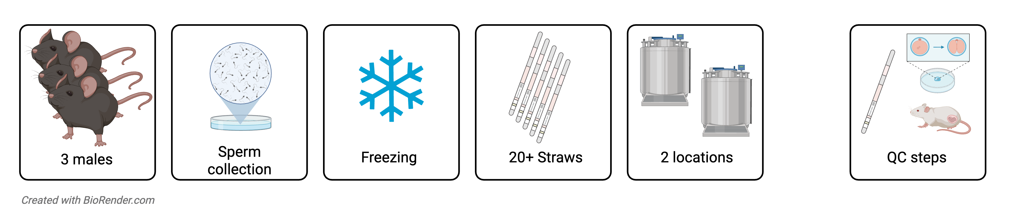
Description: Sperm is retrieved from the epididymal tissues of 3 male mice and is cryopreserved in 20 to 30 straws that are stored in liquid-phase, liquid nitrogen across two tanks in two separate locations (SRD and CCDD), to ensure sample safety and mitigate risks associated to unexpected or uncontrollable events.

Material needed: 3 males, reproductively active, 12-25 weeks old

Timeline: 2-6 weeks (dependant on QC method of choice)

Considerations: this method of cryopreservation is rapid and cheap; however, it only preserves half of the genome. This method is only recommended for single mutations on a common inbred background.

Quality Control: we provide different levels of Quality Control (QC) for different price ranges.

  1. Test thaw QC: we will thaw 1 straw the day after cryopreservation and visually assess motility and viability of the recovered sperm
  2. IVF and culture to blastocyst QC: we highly recommend this QC step. In addition to test thaw, we will also perform IVF and culture embryos up to blastocyst stage. We will provide the investigator with a fertility rate (%) for the recovered sperm. We will charge an extra cost to cover the IVF procedure.
  3. IVF and embryo transfer QC: In addition to test thaw, we will perform IVF and transfer 2-cell embryos into up to 3 pseudopregnant females to generate viable embryos/live pups. We will charge an extra cost to cover the IVF and embryo transfer procedures.

    Please note that we require you to provide your genotyping protocol, as well as full detail of the genetic content of each strain that you submit for cryopreservation.

Diagram of Sperm Cryopreservation

Embryo Cryopreservation:

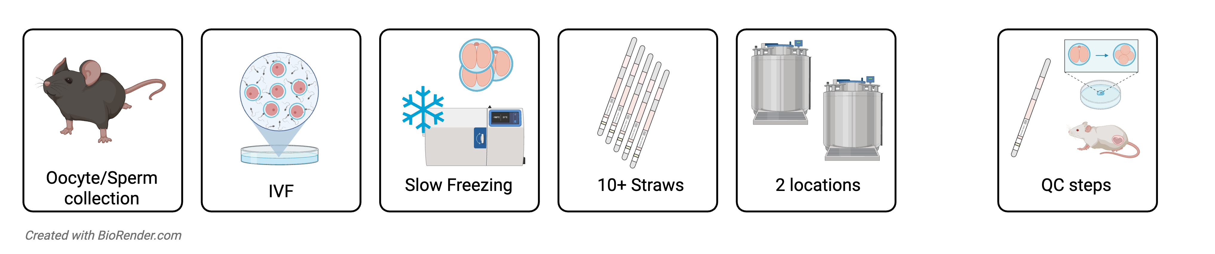
Description: Female mice are hormonally superovulated and oocytes are retrieved for in vitro fertilisation (IVF) with sperm from donor male. Resulting embryos are placed in cryoprotectant and loaded into multiple straws, which are gradually cooled and stored in liquid-phase liquid nitrogen in two separate tanks.

Material needed: Donor male and 8-10 donor females

Timeline: 12-15 weeks

Diagram of Embryo Cryopreservation

Embryonic Stem Cells Cryopreservation:

Not available, yet.

Oocyte Cryopreservation:

Not available, yet.

Cryostorage:

If you have cryopreserved mouse sperm/embryo/oocytes at another institution, we can cryostorage your samples for an annual fee. We do require that the investigator takes charge of shipping costs into our facility, and that thawing and genotyping protocols are submitted to the CIVM.

The CIVM stores all samples in liquid-phase liquid nitrogen tanks (CryoPlus1, ThermoFisher Scientific). Material retrieved from each strain is split between 2 tanks, a main and a backup tank, for redundancy. For additional safety, these 2 tanks are located in two separate buildings at ICR Sutton. Both tanks are continuously monitored by T-scan alarm systems and undergo annual service, as well as daily visual inspections.


Sperm Cryorecovery:

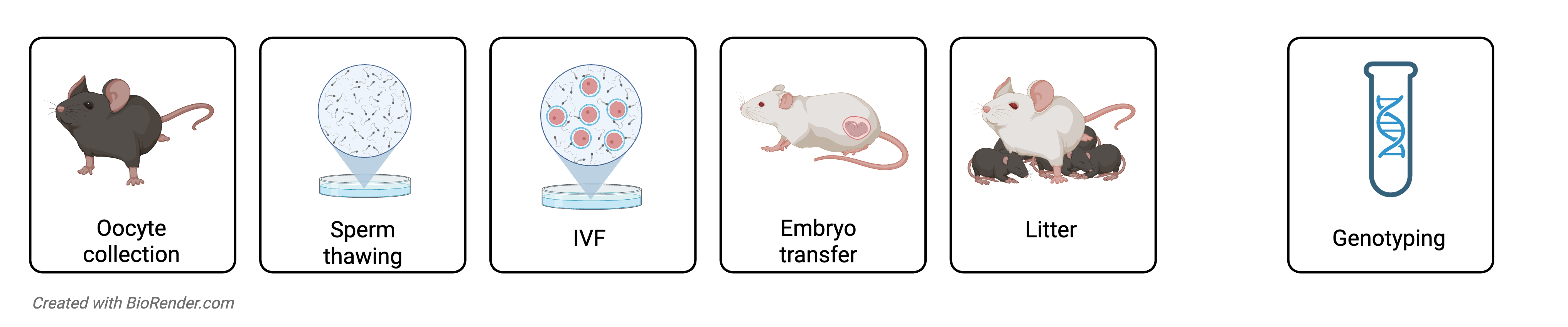
Description: Frozen sperm is cryorecovered by IVF, followed by embryo transfer. We can purchase wild-type female oocyte donors of the same genetic background, or alternatively the investigator can provide homozygous oocyte donors of the same strain.

Material needed: straw with frozen sperm and 8 to 12 females for IVF, 7-16 weeks old.

Timeline: 12-15 weeks

Diagram of Sperm Cryorecovery


Embryo Cryorecovery:

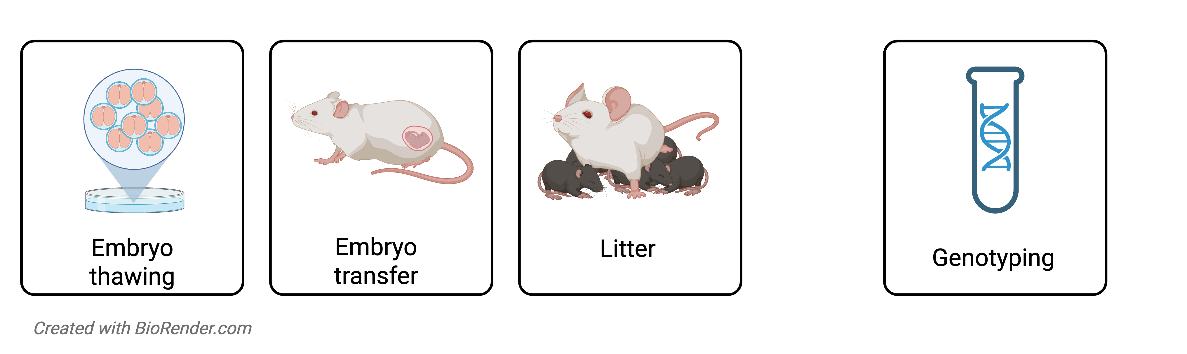
Description: Frozen 2-cell embryos are thawed and transferred into pseudopregnant females.

Material needed: straw(s) with frozen 2-cell embryos

Timeline: 8-10 week


Oocyte Cryorecovery:

Not available, yet.


Mouse rederivation

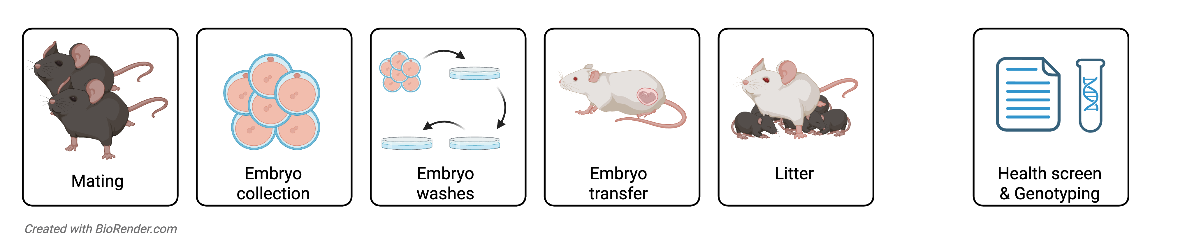
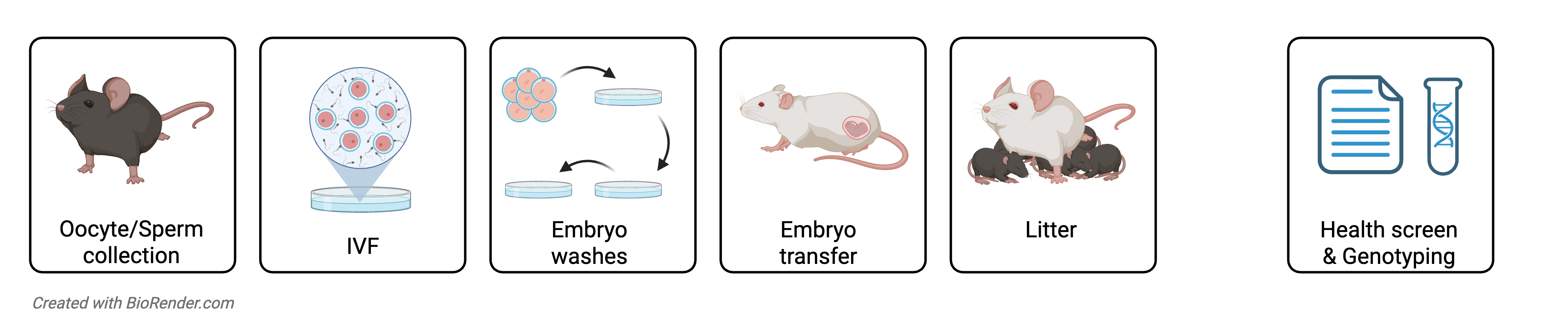
Description: Mouse rederivation is a process used to produce pathogen-free mouse colonies by removing microbial contaminants from existing lines. The procedure can be performed either through natural mating or in vitro fertilization (IVF):

  • In natural mating, embryos are obtained from donor mice and transferred into pathogen-free recipient females.
  • In IVF-based rederivation, fertilized embryos are created in vitro using gametes from donor mice and then implanted into clean recipient females.

Both methods effectively eliminate pathogens, allowing safe importation of mouse strains from lower health-status facilities into the ICR BSU. Samples from both litter and recipient mother will be sent for Health Screening and the associated costs will be charged separately to the Investigator.

Material needed: For IVF-based rederivation we require the investigator to provide 2 males, reproductively active, 12-25 weeks old, and the CIVM will purchase wild-type female egg-donors. Alternatively, if maintaining homozygosity is essential, the investigator will need to provide additional 6-10 females, 7-16 weeks old.

Timeline: 12-15 week

Mouse Rederivation Mating Diagram

Mouse Rederivation IVF diagram

We are currently setting up CRISPR/Cas9-based gene editing protocols. Soon, you’ll be able to apply for projects that involve developing new alleles based on:

  • Knockout by indel formation
  • Knockout by precise deletion
  • Conditional knockout
  • Knock-in of point mutations
  • Knock-in of small tags
  • Large knock-in
  • Exon replacement

These alleles will be developed based on Electroporation of Microinjection of CRISPR/Cas9 system reagents.

We will collaborate with you to design the best strategy and help you generate the genetically engineered mice you need for your project. 

We also provide:

  • Development of humanised mouse strains
  • Development of Patient-derived xenografts (PDX) and organoid models

Latest ICR News

08/07/26

Scientists have developed a powerful new gene-editing platform that allows them, for the first time, to systematically test the function of key proteins in mammalian cells.

Using the technology, researchers have created the first comprehensive functional map of key sites on proteins called histones, which act as spools around which DNA is wound. Histones help package the genome and play a crucial role in controlling which genes are switched on or off.  

By revealing which of these key sites are essential for healthy cells, this research – led by scientists at The Institute of Cancer Research, London – could help scientists better understand cancer.

Funding for the research was provided by UK Research and Innovation, the Royal Society and The Institute of Cancer Research (ICR), which is both a research institute and a charity. The findings were published in Nature Genetics.

Overcoming a long-standing obstacle

Small chemical tags attached to histones can alter gene activity, influence cell identity and help maintain genome integrity. Although scientists have identified more than 100 different histone modifications, understanding exactly what many of them do has proven remarkably difficult.

One major obstacle has been the complexity of mammalian genomes. Histone genes exist in many near-identical copies scattered throughout the genome, making it extremely challenging to alter every copy of a specific histone site and observe the consequences. Previous approaches often introduced unintended changes and lacked the precision required for systematic studies.

The new study overcame these limitations. The researchers developed a high-throughput CRISPR prime-editing platform capable of precisely editing all copies of mammalian histone H3 genes in their natural genomic locations. The system allows scientists to introduce targeted mutations, reverse them if required and even combine multiple mutations to investigate how different histone modifications interact.

Discovering which histone sites matter most

To demonstrate the power of the platform, the team systematically altered every lysine residue within histone H3. Lysines are amino acids that frequently carry regulatory chemical modifications, making them key control points for gene activity.

The researchers replaced individual lysines with another amino acid, arginine, preventing the usual chemical modifications from occurring. By comparing these mutant cells with carefully matched control cells, they were able to measure the contribution of each lysine to cell fitness and function.

The experiments revealed a small group of particularly important residues. Mutations affecting H3K4, H3K9, H3K14, H3K18 and H3K79 reduced cellular fitness, suggesting that the normal modifications occurring at these sites are essential for healthy cell growth and survival. Some mutations were so poorly tolerated that researchers struggled to create fully edited cell lines because cells carrying too many mutations were outcompeted by healthier cells.

In contrast, mutations at several other lysine sites had little effect on cellular fitness under the conditions tested. This suggests that histone H3 lysines make different contributions to cell function and that some may play roles in specific biological contexts that were not captured in this study. Together, the findings provide one of the clearest pictures so far of how individual histone H3 residues contribute to mammalian cell function.

The platform also allowed the researchers to study combinations of mutations. They found that some pairs of mutations had much stronger effects than either mutation alone. In particular, simultaneous mutations at positions H3K27 and H3K36 impaired stem-cell self-renewal and altered patterns of gene expression, revealing previously hidden interactions between chromatin-regulating pathways.

One of the study's notable findings involved H3K56, a histone site previously linked to DNA damage responses in yeast and fruit flies. The researchers showed that mammalian cells carrying H3K56 mutations were more sensitive to DNA damage, providing evidence that this genome-protective function has been conserved through evolution.

Laying the foundations for future cancer research

First author Daniel Price, Higher Scientific Officer at the ICR, said: “Our study establishes a scalable framework for functional interrogation of histone alterations in mammalian cells.

“The work is fundamentally a basic-science advance rather than a medical breakthrough, but the implications could be significant in the long term. Abnormal chromatin regulation is a hallmark of many cancers, and large-scale sequencing studies have identified recurrent mutations in histones and chromatin-regulating proteins across numerous tumour types.

“Although the clinical impact is not immediate, the technology provides a powerful new way to investigate how chromatin regulation contributes to cancer and other diseases, which may eventually inform future treatments.”

Senior author Dr Alex Radzisheuskaya, Group Leader of the Chromatin Biology Group at the ICR, said: “Our findings provide an unprecedented view of the histone residues that regulate mammalian genomes, and they offer researchers a powerful new toolkit for decoding the biology of chromatin.

“By finally making it possible to systematically test the function of individual histone residues, we have opened the door to a deeper understanding of how cells maintain their identity and protect their DNA, as well as how these processes go wrong in diseases such as cancer.

“We hope that both the methodology and the collection of precisely engineered cell lines generated during this project will become a valuable resource for scientists studying gene regulation, genome stability, development, cancer and epigenetic therapies.”

Image credit:  Anja from Pixabay (modified)