The competition
Five images were shortlisted for this year’s competition, showcasing the eye-catching discovery science being carried out at the ICR.
Capturing cellular processes in action is key to making new discoveries or deepening our understanding of clinical problems affecting patients, and the shortlist represents some of the very best examples of our discovery science across the ICR.
Our annual imaging competition celebrates the incredible research by our scientists at the ICR and The Royal Marsden, and the arresting images they can produce.
The selection chosen for the judges’ shortlist covers a range of cancer research fields and scientific techniques – including tumour organoids, immunofluorescence imaging and novel cancer diagnostics – to tell a story about our science with striking effect.
At the close of our competition, our shortlisted images had received 1185 interactions across all our social media channels.
Judges winner
- Rose Foster, PhD Student in the Prostate and Bladder Cancer Team in the Division of Radiotherapy and Imaging.

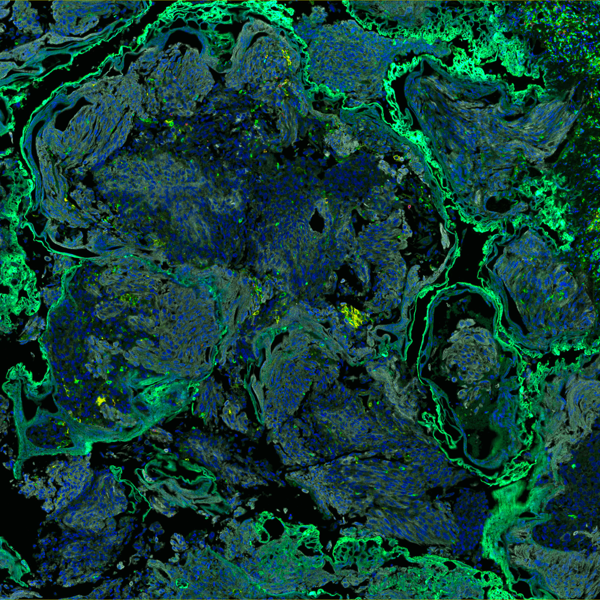
Image: Bladder cancer tumour tissue stained with fluorescent dyes to highlight proteins expressed on the cells by Rose Foster, PhD Student – Prostate and Bladder Cancer Team, Department of Radiotherapy and Imaging
Islands - Immunofluorescence staining of bladder tumour tissue
The image shows bladder cancer tumour tissue at the cellular level, stained with fluorescent dyes of different colours highlighting different proteins expressed on the cells. Tumour areas in grey and blue are surrounded by exhausted tumour-fighting immune cells called tumour infiltrating lymphocytes (TILs) in green that can’t access deeper tumour areas.
Therapeutic response for many tumours, including bladder cancer, is often linked with the number of TILs present, but the complex spatial relationship between the tumour and immune cells indicates the limitations of assessing tumours according to TIL numbers, and highlights the need for complex spatial profiling to understand the immunobiology of the tumour microenvironment.
The judges thought this entry was one of the most striking images submitted to this year’s competition that it looked like a landscape taken from above. The judges noted that the description was helpful in understanding what the image depicts.
PhD student Rose Foster said: “Entering the ICR Science and Medical Imaging competition has been an excellent opportunity to share our work with friends and family of researchers and patients in a gripping and impactful way.
“This image is the first of many bladder cancer tumours from exceptionally curated clinical trial samples that we will be imaging using multicolour immunofluorescence. My thanks goes out to all the patients providing these samples that will help us understand the intricate relationship between bladder tumours and the immune system to improve clinical decision making.”
Supporters favourite
- Melina Beykou, PhD student, Dynamical Cell Systems Team, Division of Cancer Biology and Lewis Keeble, Imperial College, London

Image: An electron microscopy image of a tumour cell attached to a lab-on-a-chip device by Melina Beykou, PhD student, Dynamical Cell Systems Team, Division of Cancer Biology and Lewis Keeble, Imperial College, London
A triple-negative breast cancer cell on a lab-on-a-chip device
A breast cancer cell attached to a microchip that can measure pH change. Cancer cells are known to acidify their tumour microenvironment, so measuring acidity levels through pH changes could be a novel way to assess cancer in the clinic.
Extracellular acidification of the tumour microenvironment plays a key role in tumour growth, metastasis, and therapy resistance. The researchers are assessing the lab-on-chip device which is made up of thousands of sensors to detect pH changes produced by cancer cell metabolism, known as the Warburg effect. Their project is working towards adapting the current technology to detect tumour acidification, which could lead to a new handheld clinical diagnostic tool for cancer.
This image received 683 likes and shares across the ICR's social media channels to be crowned our supporters’ favourite.
PhD Melina Beykou said: “It is an honour to have been shortlisted amongst some very impactful images in this competition. We are particularly excited to see the public’s reaction to our ongoing research and would like to thank everybody for their support. I would especially like to thank, PhD student Lewis Keeble, for his help in the clean room and advice throughout this project.
“We hope that this competition has given the public a little insight into some of the amazing interdisciplinary projects scientists at the ICR-Imperial CRUK Convergence Science Centre are contributing to. Our lab-on-chip technology could pave the way for the use of a hand-held diagnostic device in cancer detection."
Special commendation from our Judging Panel
Our competition judges also wanted to recognise one of our shortlisted images with a special commendation, to acknowledge the technical ability, scientific merit and originality of this entry:
- Alexia Martin, PhD student, Target Validation and DNA Damage Response Team, Division of Breast Cancer Research and the Cancer Research UK Convergence Science Centre

Image: A high-resolution image of triple-negative breast cancer organoids undergoing mitosis cell division, captured in real-time by the dual inverted single plate illumination microscopy (diSPIM) light-sheet microscope by Alexia Martin, PhD student, Target Validation and DNA Damage Response Team, Division of Breast Cancer Research and the Cancer Research UK Convergence Science Centre
3D imaging of a patient-derived breast cancer tumour organoid in different stages of cell division
Here, you can see the DNA of cells that make up a tumour organoid of breast cancer; a 3D model of the disease where you can see some cells going through mitotic cell division. The image was taken using the cutting-edge dual inverted single plate illumination microscope (diSPIM), which can capture stunning 3D images of live tumour organoids in high detail from all angles. This allows researchers to zoom in to any cell within the organoid to watch biological processes such as mitosis in real-time.
This is the first time that mitosis has been imaged in living patient-derived organoids using the diSPIM at the ICR, and the judges felt that the technical merits of obtaining this image made it worthy of a special commendation.
In breast cancer, the chemotherapy drug paclitaxel prevents mitosis by inhibiting the disassembly of microtubules in cells. This generally leads to cell death, but cancer cells can sometimes evade immediate cell death by exiting mitosis prematurely, which may contribute to drug resistance. Being able to image mitotic defects caused by chemotherapy drugs like these can help us better understand and target them, allowing us to improve patient treatment strategies.
The other shortlisted entries are
- Dr Jarama Clucas, Postdoctoral Training Fellow – Cell Death and Immunity Team, Division of Breast Cancer Research

Image: A 3D confocal microscope image depicting the highly organised, branching mammary ductal structure of mammary cells, grown from a single mouse mammary progenitor cell in culture by Dr Jarama Clucas, Postdoctoral Training Fellow – Cell Death and Immunity Team, Division of Breast Cancer Research
Studying normal breast development helps us better understand the causes of breast cancer
This is a 3D confocal microscope image of a culture of breast tissue cells grown from a single mouse mammary stem cell, showing a snapshot of the highly organised, branching mammary ductal structure that develops as the tissue grows.
By providing breast stem cells with the perfect environment to grow, we can observe and admire how breast tissue acquires its beautifully complex branching shape – from a single cell to a mini functioning organ in a dish.
Studying healthy breast tissue in the lab not only provides insights into how cancer can develop but will also aid novel drug design that minimises side effects on surrounding healthy breast tissue during cancer therapy.
- Melina Beykou, PhD student, Dynamical Cell Systems Team, Division of Cancer Biology

Image: A triple-negative breast cancer cell showing evidence of cellular senescence from treatment with palbociclib by Melina Beykou, PhD student, Dynamical Cell Systems Team, Division of Cancer Biology
To sleep or not to sleep – the role of senescent-like cells in therapy resistance
Cellular senescence is a process where cells stop dividing and can occur in response to drug treatment. Recently, senescence has been recognised to contribute to a tumour microenvironment that helps cancer cells grow and develop resistance to treatment. But we can also use senescent cells to our advantage.
This image was taken as part of a project to characterise the senescence-like cell state which arises for some cases of Triple Negative Breast Cancer (TNBC) treated with the targeted cancer drug palbociclib. Understanding the diverse characteristics of senescent-like cells that develop as cells become resistant to therapy will be key to developing new combination therapies that can overcome resistance in breast cancer.
Thought-provoking images
The judging panel for the ICR Science and Medical Imaging Competition was made up by: Dr Basil Greber, Leader of the Structural Biology DNA Repair Team in the Division of Structural Biology; Dr Nick van As, Medical Director of The Royal Marsden; June Torrance, Photographer in Residence at The Royal Marsden; Catherine Graham, Head of Brand and Creative at the ICR; and Graham Shaw, Science Communications Officer at the ICR.
Graham Shaw said: “The theme of this year’s ICR Science and Medical Imaging competition was Discovery Science, to celebrate that the most promising, innovative new treatments are only possible through fundamental research that helps us understand the underlying biology of cancer and its weaknesses. The entries this year demonstrate the broad range of discovery science our researchers are carrying out every day, and the amazing imagery that it can produce.”
Cutting-edge research
These eye-catching images illustrate just some of the cutting-edge research being carried out at the ICR, taken using sophisticated equipment purchased thanks to generous donations from our supporters.
From images like these our researchers are gaining unprecedented insights into the mechanisms that drive cancer, and new ways to target the disease to help treat patients.