Division of Structural Biology
The Division of Structural Biology aims to describe the structural and biochemical properties of proteins and the complexes they form, in order to understand the significance of these proteins in the development and treatment of cancer.
Aims and activity in this division
Researchers in the division use a variety of biochemical and biophysical techniques to understand protein structures, with a particular focus on X-ray crystallography and electron microscopy.
By combining structural biology with biochemistry and functional studies, researchers are able to gain an understanding of important biochemical interactions in the spread of cancer throughout a patient’s body.
Consequently, several group leaders in Structural Biology have joint appointments with other divisions (e.g. Cancer Biology and Cancer Therapeutics) to facilitate the exploitation of the molecular understanding of biological mechanisms in the development of new cancer therapies.
Current research activities include studying key cancer stem cell signalling processes, the role of the proteasome and the Cop9 signalosome in protein degradation and turnover, and transcription regulation.
All of these research areas have the potential to open up novel therapeutic strategies. The division also uses high-throughput screening on a variety of cancer targets, in order to identify and develop potential new candidate drugs for cancer therapy.
Facilities
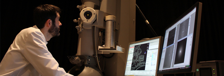
Electron Microscopy Facility
The Electron Microscopy Facility in Chelsea brings together a combination of instrumentation and expertise that supports the determination of three-dimensional protein complexes structures by cryo-electron microscopy (cryo-EM).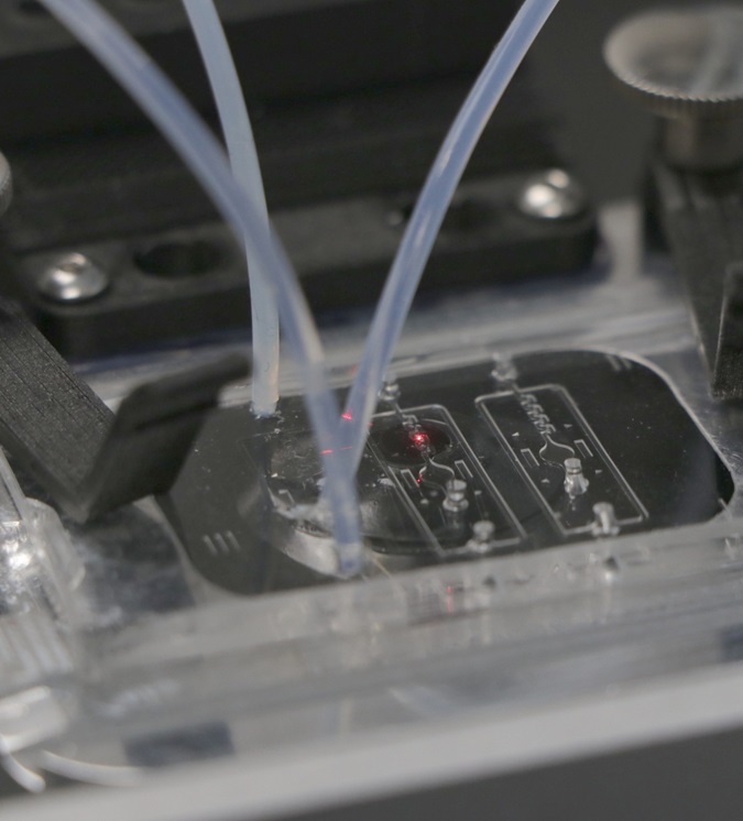
X-ray Crystallography Facility
The X-ray Crystallography Facility provides tools for the crystallisation and X-ray analysis of biological material.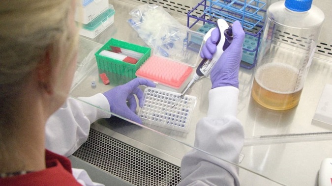
Baculovirus Facility
The fully equipped Baculovirus Facility provides a means of expressing proteins and protein complexes using a Baculovirus / insect cell system (BICS).-lab.jpg?sfvrsn=e525f31_1)
Correlative Light and Electron Microscopy (CLEM) Lab
The CLEM lab is jointly run by researchers from the Electron Microscopy and Light Microscopy facilities. We provide access to equipment, training and help to design custom workflows for in situ structural biology projects.
Chelsea Biophysics Facility
The biophysical equipment provided by the facility allows researchers a wide range of options for characterising their biological samples.Research group leaders
.jpg?sfvrsn=4c485212_1 "Sebastian_Guettler_2 (1)")
Professor Sebastian Guettler
Head of Division
Structural Biology of Cell Signalling.tmb-propic-md.jpg?Culture=en&sfvrsn=d41885a5_9 "Laurence Pearl (Profile)")


