
A special device for holding mice while they undergo magnetic resonance imaging (MRI) scans on clinical MRI machines – those used on humans – can enhance imaging investigations in mouse models of neuroblastoma, researchers have found.
The device, known as a ‘mouse hotel’ by its manufacturer, contains three bays for the mice, and a radiofrequency coil that enables the MRI scanner to produce high-quality images from multiple mice.
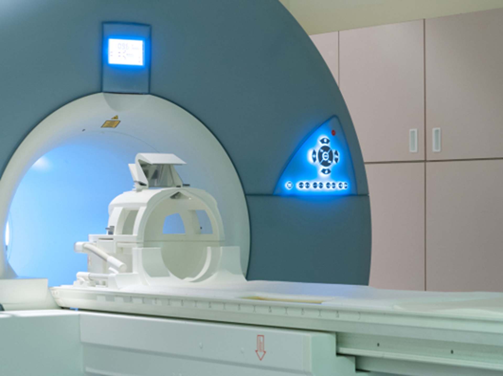
Most research in mice uses small, specialised scanners, but the ability to use clinical MRI systems brings a number of benefits – including better soft tissue contrast between healthy and cancerous cells, and the ability to simultaneously examine several mice.
It also makes the research more directly comparable to human MRI investigations.
Researchers are studying neuroblastoma – a childhood cancer of the nervous system – using mice in which tumours spontaneously arise within the abdomen. Cancers in these mice closely mimic human neuroblastomas, but require improved non-invasive imaging methods to more accurately assess tumour growth and response to treatment.
The study was supported by a range of organisations including Cancer Research UK, the Department of Health, and the NIHR Biomedical Research Centre at The Royal Marsden NHS Foundation Trust and The Institute of Cancer Research, London.
Research at the ICR is underpinned by generous contributions from our supporters. Find out more about how you can contribute to our mission to make the discoveries to defeat cancer.
Comparing both systems
Writing in the British Journal of Cancer, scientists from The Institute of Cancer Research explain how they compared the ‘mouse hotel’ system to a traditional small-scale MRI.
Mice with neuroblastomas were scanned on both systems within 24 hours, and the results were analysed side-by-side.
The researchers were able successfully to use the clinical MRI system to scan three mice at the same time, with good enough resolution to assess their tumours, and found no significant difference between the two systems for measuring tumour volume.
They also saw strong similarities for more technical measurements of the mouse tumours, which can indicate the state of their blood supply and the extent of cell death. These measurements are important, as they can be used to assess how well a tumour is responding to treatment.
Finally, the team were able to assess tumour response to chemotherapy in the mice using the clinical MRI platform.
Best of both worlds
Study leader Dr Simon Robinson, Team Leader in Magnetic Resonance at the ICR, said:
“Clinical MRI scanners are increasingly being used for animal studies because they allow us to make more accurate comparisons with research in patients.
“Our research shows that using a ‘mouse hotel’ allows us to get the benefits of a clinical MRI scanner at the same time as the image quality of a pre-clinical system.
“In this way, obtaining simultaneous imaging data from multiple mice provides a high-throughput approach for evaluating new treatments for neuroblastoma.
“In addition to the impact of non-invasive imaging on reducing the number of animals used in our research, the clinical relevance that comes with imaging mice on a clinical scanner also represents a refined study design that aligns with the principles of the ‘3R’s’ – replacing, refining and reducing – in animal research, which the ICR strongly supports.”