An artificial intelligence (AI) that reconstructs MRI images of moving tumours can do so in seconds, offering a significant improvement on current methods to optimise radiotherapy treatment in the clinic.
The AI, called Dracula (short for ‘deep radial convolutional neural network’) can replicate four-dimensional MRI images of a patient’s whole anatomy, including tumours and healthy organs, from low quality images containing artefacts – visual anomalies not present in reality that arise from scans with incomplete information.
It can take up to several hours to reconstruct high quality 4D MRI images from these under-sampled scans, whereas the AI takes an average of 28 seconds.
These reconstructed images could accurately guide the delivery of radiotherapy to tumours by tracking their movement while a patient is breathing. The process is called magnetic resonance guided radiotherapy (MRgRT) and allows radiotherapy treatment to be adapted accordingly to improve patient outcomes.
The research was published in the journal Radiotherapy and Oncology, and was led by scientists at The Institute of Cancer Research, London and The Royal Marsden NHS Foundation Trust. It was funded by Cancer Research UK, NVIDIA and the NIHR Biomedical Research Centre at The Royal Marsden and the ICR.
Faster image acquisition
To treat patients with tumours that move as they breathe, 4D MRI images are needed to show the 3D volume of the tumour and surrounding organs at different time points during breathing – known as a respiratory phase.
By combining numerous respiratory phases, the midposition image can then be calculated from the 4D MRI image. Midposition images reveal the average position of the tumour and its movement from breathing, and are needed to accurately plan radiotherapy treatment.
An alternative to treating these types of tumours is to have a patient hold their breath so the tumour no longer moves, but this can be strenuous for the patient and makes treatment longer and more difficult.
Dracula allows 4D MRI and midposition images to be obtained in seconds – and potentially online just before treatment – to guide the delivery of radiotherapy in real-time, as well as make treatment more adaptable based on the patient’s anatomy.
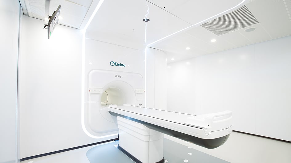
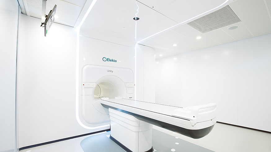
In the clinic, the images reconstructed by Dracula could be used to guide the ICR and The Royal Marsden’s MR Linac machine – a novel combination technology that uses an MRI scanner and linear accelerator to locate and deliver radiotherapy doses to tumours. This means high hits of radiation could be precisely targeted to just the tumour, minimising the risk of affecting healthy organs.
Clinically acceptable
In line with its name ‘neural network’, the AI works like a simplified version of neurons in the brain and learns to reconstruct higher quality 4D MRI images via a series of training examples. The researchers used low quality 4D MRI images that are unfit for clinical use as the input data for Dracula.
With both information on the spatial dimensions and respiratory phases of a patient’s tumour and surrounding organs, the researchers could train Dracula to produce 4D MRI and midposition images that were considered acceptable for use in the clinic by both a radiologist and a radiation oncologist.
Dracula’s performance was also verified against 4D MRI images reconstructed by another algorithm, MoCo-HDTV, for comparison, with image quality graded on a five-point Likert scale – zero denoting unreadable and five excellent.
Dracula-reconstructed images received scores between 1.8 and 3.4, with the experts reporting some minor blurring and streaking that reduced visibility.
Though not scoring as high as images produced by the time-intensive MoCo-HDTV, Dracula was still able to visualise the patient’s tumour and at-risk organs – in this case the heart and oesophagus – very well, so that it could be used in practice to plan and guide radiotherapy treatment.
Study leader Dr Andreas Wetscherek, Team Leader in Magnetic Resonance Imaging in Radiotherapy at the ICR, said:
“Using an AI to rapidly reconstruct 4D MRI images of a cancer patient’s anatomy lets us accurately determine the location of tumours and characterise their motion right before radiotherapy treatment, which will enable us to increase the radiation dose targeting just the tumour and not healthy organs.
“This study demonstrates the potential of neural networks to achieve comparable imaging results to traditional methods in only a few seconds. It would be especially useful for cancers where we need to avoid the healthy organs around the tumour, such as pancreatic cancer, which currently limits the delivery of high doses of radiation to the tumour.”
