Our research into cervical cancer
We have made waves when it comes to cervical cancer. Our scientists have worked on improving imaging technology that helps guide decision-making in surgery, and helped trial new drugs in patients with cervical cancer as part of clinical trials with our partners at The Royal Marsden hospital.
We helped unravel the puzzle of how the Human Papilloma Virus (HPV) causes cervical cancer and published the first data showing that testing for HPV reduces the number of women who go on to need a smear test down the line.
Header image: Microscope image of squamous cell carcinoma, Image credit: National Cancer Institute
Our progress against cervical cancer
Ultrasound
Research led by Dr Emma Harris and Dr Susan Lalondrelle at the ICR and The Royal Marsden has looked at pairing computerised tomography (CT) scans with ultrasound imaging to produce more detailed images that allow clinicians to better plan treatment for cervical cancers.
CT scans are used to plan where radiotherapy will be applied, but it’s tricky to get this just right with cervical cancer. Natural movements of the bladder and bowel mean that the location of the tumour can change rapidly, leading to unpleasant side effects for some women.
Combining ultrasound and CT scans is allowing clinicians to better outline positions of the uterus and makes treatment much easier to plan and administer safely.
Clinical trials
Researchers at the ICR and The Royal Marsden are trialling new drugs in women with gynaecological cancers including cervical cancer. In 2019, scientists published results from a global clinical trial of nearly 150 patients with a variety of cancer types who had stopped responding to standard treatments, including patients with cervical cancer.
The new drug being trialled, tisotumab vedotin (or TV for short), releases a toxic substance to kill cancer cells from within, and the trial showed that over a quarter of patients with cervical tumours responded to the treatment. Professor Johann De Bono and his team led the trial, and work on the drug is ongoing.
MRI
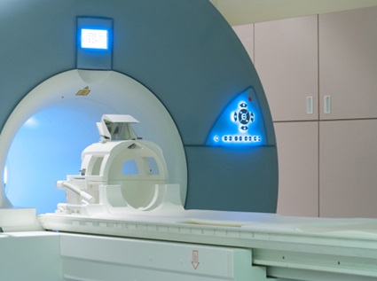
The ICR led on research which showed that a specialist type of magnetic resonance imaging (MRI) test could limit the extent of surgery needed in younger women with early-stage cervical cancer.
Professor Nandita deSouza and her group developed a new MRI scan that produces a greater resolution and contrast between tumour tissue and normal tissue, giving more detailed information about the extent of the disease in the cervix.
This means that some women can be spared radical hysterectomies and radiotherapy, both of which come at cost of a woman’s fertility. The scans allow clinicians to preserve more of the cervix in some women, and leave the uterus in place, meaning women can still become pregnant and deliver by caesarean section.
Cervical cancer clinical trials
In our Clinical Trials and Statistics Unit (ICR-CTSU), we translate cutting-edge science into quality clinical trials that can transform cancer care.
Here are the latest trials that include patients with cervical cancer. If you or someone you know is a cervical cancer patient, then you may be able to support our research by taking part in a trial that’s “Open to recruitment”. Or see all clinical trials.
Your support helps
Please donate today to help more people survive cervical cancer. As a UK charity, our life-saving research relies on the generosity of individuals and organisations. Our supporters help us make a difference to the lives of cancer patients and their families everywhere.
Cervical cancer discoveries

New ‘Trojan horse’ cancer treatment shows early promise in multiple tumour types

Imaging probe outperforms the best external detector for early cervical cancer

Cervical Cancer Risk for Women over 50

.tmb-hbdesktop.jpg?Culture=en&sfvrsn=6ac42851_1)