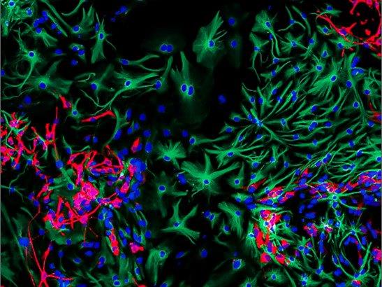

Image: The winning image by PhD student Sumana Shrestha, showing differentiating brain cancer cells used to study glioblastoma
PhD student Sumana Shrestha at The Institute of Cancer Research, London, has won the ICR’s Science and Medical imaging competition with a colourful image of neural stem cells to study glioblastoma, as well as the public vote, with 212 votes for her detailed image of microparticles.
Researchers from the ICR and The Royal Marsden submitted some fantastic images to this year’s competition, with a wide array of images that tell vivid stories about our science.
Entries to the competition have all been created in the course of our pioneering cancer research – but at times they can look like a piece of modern art, and they each tell a compelling story about our research.
The most engaging and exciting image selected by our panel of judges is named the winner of the ICR Science and Medical Imaging Competition.
Our followers on social media picked the supporters' favourite through a public vote, with over 800 votes submitted across Facebook, Instagram and Twitter.
Judges’ winner: Differentiating brain cancer cells by PhD student Sumana Shrestha
The winning image was taken using a technique called confocal microscopy and shows neural stem cells from mice which are being used to study glioblastoma, an aggressive type of brain cancer. The cells were genetically modified and exposed to a virus to develop glioblastoma, before mice were treated with microRNA repressors and activators to change how the cells grow in the brain and specialise.
A process called immunostaining shows up a garden of stem cells growing and specialising into specific types of cell, to carry out jobs in the brain. Star shaped cells called astrocytes are marked in green, which perform a range of functions in the brain, while neurons are marked in red, cells in the nervous system that help transmit information.
MicroRNAs are proteins in cells that help regulate gene expression, and in cancer they can be severely malfunctioning. Modulating microRNA levels in brain cancer cells could have a major impact on how the tumour cells develop, which could help to make the disease less aggressive and easier to treat.
The judges picked this as their wining image noting the striking use of colours in the image, and how Sumana has used her image to illustrate how her research is helping to tackle cancer.
Catherine Graham, Head of Brand and Creative at the ICR, said:
"Sumana’s image is really eye-catching and it is a great example of the technical skill and creative flair of our researchers, and the visual impact that can be captured even in a disease like cancer.”
“Choosing the winning image from all of those submitted to the competition this year was challenging, but it was a pleasure to see the variety of our researchers’ work and to learn more about their science.”
PhD student Sumana Shrestha said:
“This is very unexpected news for me as the competition was so strong, but I am over the moon to find that my image has won! Many thanks to the judges for taking their time to go through the images. I am very excited for the announcement, and again, overjoyed about the results.”
Supporters Favourite: Dimpled ‘golf ball-like’ microparticles by PhD student Sumana Shrestha

The image shows microparticles, tiny beads about the width of a human hair, taken using scanning electron microscopy. To image the microparticles, they are first coated in a layer of gold just nanometres thick to make them conductive. The particles have been etched using an antibiotic called fusidic acid to create dimples on their surface like a golf-ball.
Microparticles can be used to deliver drugs in the body, but studies have shown that the textured surface of microparticles can also have an effect on cell behaviour.
Cells are influenced by the topographical features of their surroundings, and the 'dimples' on the treated microparticles provide an environment that can be used to control proliferation or differentiation of stem cells. Manipulating fundamental cellular processes using microparticles could have huge implications in cancer, as well as applications in regenerative medicine.
This image received a total of 212 across the ICR’s social media channels to be crowned our supporters favourite, with an ICR follower on social media thinking the microparticles looked like pollen grains.
Science Communications Officer Graham Shaw, who organised the competition, said: “It’s surprising that two submissions from one researcher have won both prizes in the competition, but this black and white image clearly captured the imaginations of our supporters on social media.
“The detail on the microparticles is amazing, and their potential to alter cells as a way to treat cancer is a really powerful idea. This image helps to convey how this has been implemented in cancer research.“
Our public shortlist
Eight of the most highly rated and vibrant images from this year's competition were voted on by our supporters for their favourite image. Learn more about the other amazing images that made it onto our shortlist.
The eye of the Storm: the tumultuous yet spectacular nature of cancer tumours by Dr Lisa Pickard and Louise Howell

The eye of the storm: the tumultuous, dramatic nature of cancer tumours. Lab grown mini-tumours are better at capturing the unique characteristics of tumours in patients, and can be used to understand how they might respond to cancer drugs.
Melanoma on a Chip by Professor Chris Bakal and Nick Moser at Imperial College, London

Melanoma on a chip: part of a melanoma cell has been blasted away using a technique called ion beam milling. This process helps researchers look inside cells in unprecedented detail, to help them understand the processes happening inside.
Melanoma cell invasion in 3D by Vicky Bousgouni, Professor Chris Bakal and David Robertson

Invading melanoma cell in 3D. A cell from aggressive skin cancer is growing into and around fibres of collagen, mimicking how cancer invades tissue. Cancer remodels its environment to spread around the body, so understanding this process could make it easier to treat.
Super-resolution microscopy image of cell focal adhesions by PhD Student Ian Jones, Dr Lucas Dent, and Professor Chris Bakal

The first ever super-resolution microscopy image taken of focal adhesions – molecules inside cellular structures which help cancer cells move and spread around the body.
‘The Art of Deception’ by Dr Jarama Clucas

‘The Art of Deception’. These aggressive breast cancer cells look like structures normally seen in healthy breast tissue. Cancer use deception to hide from the immune system and avoid treatment, but growing samples in the lab can be used to test treatments that could help our body fight the disease.
Forward and reverse translation by PhD Student Somaieh Hedayat and Louise Howell

Tumour organoids are mini replicas of a patient’s tumours, and could be used to rapidly test a patient’s cancer against a range of cancer drugs to see which ones may be most effective.
Cutting-edge research
These eye-catching images illustrate just some of the cutting-edge research being carried out at the ICR, taken using sophisticated equipment purchased thanks to generous donations from our supporters.
From images like these our researchers are gaining unprecedented insights into the mechanisms that drive cancer, and new ways to target the disease to help treat patients.