
People have been exploring with the help of magnetic fields ever since the twelfth century, when explorers first started using compasses to guide ships on cloudy nights. The magnetic fields our researchers use today are much stronger and generated by high-tech machines, but the goal is the same – to provide direction to our explorations where we’re not able to see with our own eyes.
Magnetic Resonance Imaging (MRI) is a non-invasive technique that scans the body using powerful magnets. MRI machines used to scan humans employ magnetic fields of 1.5–3.0 Tesla (15,000–30,000 gauss) – pretty impressive considering that the earth’s magnetic field, which protects us from the damaging solar wind and has allowed humans to use compasses for navigation across the globe, only measures 0.5 gauss.
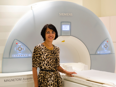
Professor Nandita deSouza is Co-Director of the Cancer Research UK Cancer Imaging Centre at The Institute of Cancer Research, London, and The Royal Marsden NHS Foundation Trust. She is using MRI imaging to transform the way we treat patients with cancer, by improving the information about tumours on which treatment decisions are made. “Imaging has the power to reveal what is inside our bodies, without having to make a single incision,” explains Professor deSouza. “But imaging the structure of tissues alone may not provide us with enough information to fully characterise or monitor tumours. By using MRI, we can perform ‘functional imaging’, and can see how tissues function and behave, providing crucial insights into the nature of cancers. We work on a range of tumour types, but I have a particular focus on prostate and gynaecological cancers, and tumour that involves bone where current techniques for diagnosis and monitoring need improvement.”
We recently covered in a press release and video a scanning technique called whole-body, diffusion-weighted MRI. Using this method, Professor deSouza and colleagues imaged a patient’s entire body, revealing where myeloma – a disease which affects plasma cells in the blood – had affected the bones. Professor deSouza says: “Using this new type of MRI scan, we could improve care for myeloma patients – reducing the need for bone marrow biopsies, which can be painful for the patient, and often fail to show doctors how far the disease has spread. And by looking at all the bones in the body in one scan, we are no longer relying on individual bone X-rays. We can now measure the involvement of individual bones and follow their response to treatment. What is more, the results can be visualised immediately, so we can see straight away where the cancer is and measure its extent. Doctors can use these scans to make more informed decisions. If a treatment isn’t working, the patient can be moved onto new therapies that might be more effective much more quickly.”
Using MRI techniques, Professor deSouza has also successfully identified imaging measurements in cancers that present early. Professor deSouza says: “We have managed to predict men whose early prostate cancer will progress and require treatment. This has an enormous impact on decision making about who to treat, avoiding unnecessary treatment and the radical removal of the prostate. For cervical cancer – another tumour that presents early and predominantly affects women under the age of 45 – surgical treatment is necessary. By using a probe in the body to achieve high resolution images that provide this information, we can help experienced surgeons decide whether or not they could safely limit their surgery and enable the woman to retain fertility.
In other tumours such as ovarian cancer, disease is detected late, and the tumour has already spread. Professor deSouza explains: “At this stage, the cancer is much more difficult to treat and patients build up resistance to treatment, so we are always looking for ways to identify non-responsive patients early in the course of treatment. Using functional MRI, we potentially can identify within a month of treatment those patients likely to benefit from the full 18-week course of chemotherapy. In future, this will enable earlier decisions on changing treatment and spare the patient from weeks of potentially ineffective treatments with unpleasant side-effects. It also gives us a visual picture of the distribution of the tumour. Most importantly, we can identify individual tumour deposits that are not responding to conventional treatment for which other treatment options – including new drugs specifically targeted to the tumour type or surgical removal – can be considered. We ultimately want to integrate diagnostic techniques with minimally invasive treatments for cancer – and it’s something that we are making real headway on.”
Functional imaging provides a means of truly delivering personalised medicine. Researchers can non-invasively detect, diagnose, characterise and monitor tumours before and after therapy, guiding doctors to select and plan individualised treatments and sparing patients needless and ineffective therapies. Like a compass magnet, the powerful magnets used for MRI are providing direction in the fight against cancer.