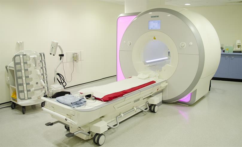
Image: An imaging suite courtesy of Joe Dunckley for the ICR, 2014
A honeycomb of tungsten built using 3D printing could help take better quality images of processes inside the body, a new study shows.
Researchers at The Institute of Cancer Research and our partner hospital, The Royal Marsden, tested a new component made using 3D printing for gamma cameras, which are used to take images of radiation emitted by pharmaceuticals injected into cancer patients.
The new component is a shield made of tungsten, a rare metal. It could help gamma cameras produce more detailed images than with the lead shields that are currently used.
In tests of the new shield, the results of which were published in the European Journal of Nuclear Medical Imaging, tungsten performed better than lead by blocking more unwanted radiation.
Taking images using gamma rays
Gamma rays are powerful beams of radiation emitted by the breakdown of the nuclei of radioactive atoms.
Scientists use gamma cameras to map processes of organs in the body, creating images from the gamma rays emitted by injected radioactive materials.
A crucial element of gamma cameras is a dense shield, or collimator, usually made of lead. The shield has a honeycomb structure of holes to allow enough radiation through to create an image, while blocking unwanted radiation so the image is sharp.
However, the relatively low density of lead affects the design of collimators that can still produce acceptable images, limiting the detail that can be seen using gamma ray cameras.
Research at the ICR is underpinned by generous contributions from our supporters. You can contribute to our mission to make the discoveries to defeat cancer.
Scientists at the ICR investigated if a shield made of a denser metal, like tungsten, could do a better job by blocking radiation more effectively.
In collaboration with Wolfmet of M&I Materials, a company in Manchester, they tested 3D printed tungsten, which demonstrated improved properties to that used in a typical lead collimator.
The tungsten shield can produce higher resolution images of organs with gamma cameras than lead shields. It could help researchers see processes in the body in greater detail.
The study was supported by the NIHR Biomedical Research Centre at the ICR and The Royal Marsden.
Lead author of the study Dr Jonathan Gear, Clinical Scientist in the Joint Department of Physics at the ICR and The Royal Marsden, said:
“Gamma ray radiation is very powerful so you need heavy shielding to produce a clear image. Most gamma ray cameras use thick lead, but denser materials like tungsten could be as effective while letting more useful radiation pass through to produce higher quality images.”
“Our research shows that 3D printed tungsten could be used in gamma cameras, offering greater flexibility in the sorts of designs that could be used in the future.”