
This week is International Medical Physics Week 2021, which runs from 26-30 April and recognises the important contributions that physics has made to the field of medicine.
Medical physics is the application of physics to healthcare. When it comes to cancer research, most people are likely to think of images of X-rays and radiotherapy as the leading contribution of physics to the field.
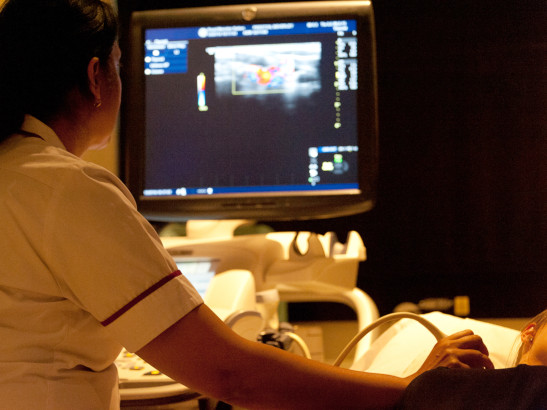
What they might be less likely to think of though is ultrasound. But the ICR has been at the forefront of developing ultrasound for medical diagnosis, and more widely in cancer research.
Dr Emma Harris and Professor Jeff Bamber both lead research programmes at the ICR relating to medical physics. They told me about some of the ground-breaking discoveries made by the ICR in ultrasound research, and new ways that ultrasound is being used to treat cancer.
Early ultrasound pioneers
Professor Jeff Bamber is leader of the Ultrasound and Optical Imaging team at the ICR, and he has been a pioneer in developing ultrasound for over 40 years.
Ultrasound uses high frequency sound waves that pass through human tissue and can be picked up by sensors to build a map of tissues and structures in the body. The most well-known example of ultrasound use is scanning expecting mothers to see their babies inside the womb.
To be useful in cancer research, scientists need to understand how the physical characteristics of tissue inside the body match with the ultrasound images they produce.
In 1979, the ICR was one of the first organisations in the world to produce computer simulations of ultrasound images. From this, Professor Bamber developed methods to remove interference from ultrasound images called ‘speckle’, which are carried out in different ways on almost every commercial ultrasound system available today.

Image: An image of a human thyroid, with ‘Adaptive speckle reduction’ applied on the right, from Bamber, J. C., & Daft, C. (1986).
These important studies helped scientists to understand the images produced by ultrasound, and use them to learn more about cancer.
Ultrasound diagnostics and monitoring treatment
From these initial steps modelling ultrasound, Professor Bamber and colleagues developed techniques to measure the physical properties of tumour tissue, including an important technique called freehand strain elastography.

Image: A freehand strain elastography image of breast cancer, that measures the stiffness of tumours compared with healthy tissue.
The tissue inside cancers can be stiffer than the surrounding healthy tissue, and their research used ultrasound to measure the stiffness of tumour tissue compared with healthy tissue. Using ultrasound elastography to measure tissue stiffness is now a common practice in medical physics.
ICR researchers are continuing to develop new ways to use ultrasound to benefit patients, such as guiding radiotherapy treatment.
Ultrasound and radiotherapy
Radiotherapy aims to focus high-energy beams of radiation at tumours to kill cancer cells. However, the natural movement of organs in our body can cause tumours to move around.
Ultrasound is a relatively cheap and easy way to look for this movement to help improve radiotherapy.
Dr Emma Harris is Leader of the Imaging for Radiotherapy Adaptation Team at the ICR, and in 2010, her research showed that ultrasound could track the movement of organs in the upper abdomen as patients as they breathed in and out. A clinical trial in 2018 of patients with prostate cancer demonstrated that ultrasound could guide radiotherapy during treatment, giving doctors second-by-second information about the location of tumours and organs, and correcting treatment accordingly.
Dr Harris’ other research interests include leading studies of a technique called spatial compounding, which could improve ultrasound images of the uterus for radiotherapy treatment.
The technique uses overlapping ultrasound scans to produce clearer images, and her team are exploring new ways it could benefit planning for radiotherapy by producing a wider field of view, allowing a larger section of the body to be imaged with higher image quality.
Shear wave elastography
ICR researchers are also developing a high-resolution form of vibrational shear wave elastography, which uses vibrations to assess the stiffness of tumour tissue.

Image: An illustration of vibrational shear wave elastography. A vibrational source (the shaker) vibrates against the skin creating shear waves which travel through the body. Ultrasound is used to image the shear waves. Shear waves move faster (and have a longer wavelength) in tumours compared with surrounding normal tissue.
They are using this ultrasound technique to understand the elastic properties of tissue in mice, to better understand how treatment effects tissue in patients.
Their research could help to develop ultrasound diagnostics for radiotherapy that shows where in tumours radiotherapy is working and where it isn’t, which could help doctors better plan treatment for patients.
The future – ultrasound therapies
Applications like shear wave elastography point to even more practical applications for ultrasound, where the waves themselves could help to treat patients’ cancers.
High Intensity Focussed Ultrasound, for example, uses highly focussed beams of ultrasound to kill cancer cells. ICR scientists have played a leading role in developing this innovative technology.
And working with partners in industry, the ICR has recently shown how an exciting new ultrasound treatment called Acoustic Cluster Therapy could help treat patients with cancer.
The technique involves injecting clusters of microbubbles and microdroplets with chemotherapy drugs, and then uses ultrasound to vibrate blood vessels inside patient’s tumours and make them more able to absorb drugs.

Image: Diagram of Acoustic Cluster Therapy releasing chemotherapy drugs into tumours using ultrasound.
Not only could Acoustic Cluster Therapy make chemotherapy more effective while reducing harmful side-effects, this painless and non-invasive technology could be used for other cancer treatments and also for other diseases.
Now the ICR is testing if the same approach can make cancer cells more sensitive to radiotherapy.
From using ultrasound to improve cancer diagnosis and assessment, to delivering better treatments with fewer side-effects, our ultrasound research has made a big difference in its use in medicine and could lead to new ways of treating cancer.