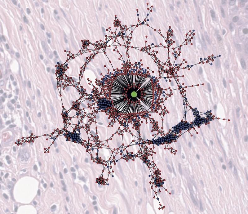

Image: A digitised image of the tumour microenviroment, showing hotspots and cold spots linked to characteristics of the cells. Image Credit: Yinyin Yuan
The field of pathology is one of the oldest in medicine and vitally important to treating cancer. Pathologists are doctors that specialise in evaluating cells, tissues, organs and test results to help diagnose patients. For people who may have cancer, they use their expertise to determine if they do and what type of cancer they have.
Advances in microscopy, imaging and DNA sequencing are uncovering more information about tumours than ever before. Now a new, high-tech specialism called digital pathology promises to make sense of all this data, and to make it easier and faster to diagnose cancer.
For National Pathology Week, we’re highlighting a specialism called digital pathology, which combines our expertise in clinical research with cutting-edge tissue analysis to help diagnose cancer.
Going digital
Around the world, pathology departments are starting to digitise pathology images and information, so that researchers and clinicians can analyse them using computational tools.
New tumour imaging systems can scan slides of tissue samples taken from patients, so that they can be shared with colleagues, and assessed alongside molecular, radiology and pathology data.
Increasingly, digital pathology aims to integrate data from different sources using AI-driven analytical tools, to help spot patterns and crucial clues that would not be visible to the naked eye. Digital pathology offers the chance to make a huge difference to the way we understand, diagnose and characterise cancer, and to help transform how cancer is treated.
Building digital pathology
We are building our expertise in digital pathology under the leadership of Professor Yinyin Yuan and Professor Manuel Salto-Tellez.
Professor Yuan leads the Computational Pathology and Integrative Genomics Team, which aims to train powerful computers to automatically identify cancer cells in pathology samples from the clinic. Professor Salto-Tellez joined us in the summer of 2020.
Their teams are leading efforts in partnership with colleagues at The Royal Marsden, our partner hospital, to ensure all our pathology images are captured digitally. By translating pathology samples into digital images, scientists can use the latest cancer tissue analysis techniques to enhance cancer diagnosis and treatment.
Digital imaging will allow doctors to extract new information from existing images, or look at multiple biological markers of cancer at the same time, while information can be accessed and analysed remotely.
Digitisation will also help our researchers to develop an array of new computational image analysis tools, using AI to support pathologists in making key decisions about a cancer diagnosis.
AI and machine learning
Computers are excellent at finding patterns in data and by using tools like AI and machine learning, they can be taught how to identify features in digital pathology images that are linked with cancer.
Professor Yuan, Professor Salto-Tellez and their colleagues at the ICR and elsewhere are building a range of laboratory and computer tools to process and classify images of tumour tissue, to increase our knowledge about the biology of cancer and help pathologists make more accurate cancer diagnoses.
Computers can mark areas with similar features within tumour samples to quickly identify cancer cells and other tissue types. These can be compared with other samples and assessments by pathologists to fine-tune the results, and to spot patterns that can’t be seen by doctors. This could help to detect cancer before symptoms occur or make assessments of whether treatments are working earlier.
By drawing on the rich information contained in these images, and combining it with other types of information such as DNA sequencing data from cancer cells, the researchers can visualise cancer and its surrounding tissues in new ways.
These new tools could help us understand how cancers interact with their environment as they develop and spread, diagnose patients more quickly or precisely, and predict how they might respond to a treatment.
Using the multitude of digital information produced when a person is scanned and tested for cancer, digital pathology could uncover amazing new information about the disease, and transform its diagnosis and treatment.
This article was adapted from Search, our twice-yearly newsletter for our supporters. Sign up to receive Search to read more about our latest news, research achievements and interviews with our world-leading scientists and clinicians.