Photoacoustic Imaging and Emission Spectroscopy of Tumour Vascularisation
A Gertsch, M Jaeger, NL Bush, JC Bamber; in collaboration with S Eccles, Section of Cancer Therapeutics; M Frenz, University of Bern, Switzerland; M Fournelle, R Lemor, Fraunhofer Institute for Biomedical Technology, Saarbrucken, Germany; L Masotti, El.En. SpA, Florence, Italy; H Wittig, tp21 GmbH, Saarbrucken, Germany
Source of funding: European Union Marie Curie and Framework 6
Even single wavelength photoacoustic imaging of the spatial distribution of blood content has considerable potential value for tumour diagnosis, prognosis and monitoring response. Photoacoustic (PA) models of large blood vessels, which assume a homogeneous optical absorption, do not provide good descriptions of tumour microvasculature. We therefore introduced the concept of a randomly inhomogeneous optical absorption into the photoacoustic imaging model, by analogy to the inhomogeneous scattering model used for many years in ultrasound imaging.
Phantoms, simulations and an analytical model have demonstrated that a degree of absorption inhomogeneity improves the quality of images of macroscopic targets, as measured by target contrast-to-noise ratio and by completeness of the displayed target boundary. At the extremes of small scale and large scale inhomogeneity, mean PA (speckle) signal intensity increases with absorber scale r as r3, and decreases as r-1, respectively. As a result, there is an intermediate scale of inhomogeneity, associated with the bandwidth of the ultrasound transducer, at which the overall target visibility is maximum.
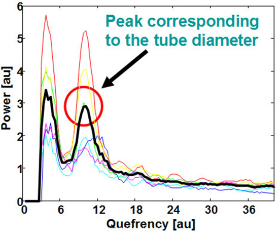
It was also shown that a cepstral analysis of the acoustic emission spectrum may be used to automatically determine the size of microscopic optical absorbers. Recent work demonstrated that, in a phantom containing multiple randomly-distributed vessels, the characteristic vessel size remained measurable by this method so long as it was substantially smaller than the mean spacing between vessels (Figure 12). Ongoing work aims to determine whether such methods can be made provide an in vivo vessel size index for eventual use as a cancer response biomarker.
 Fig.12. Averaged photoacoustic (PA) emission cepstrum from a multivessel phantom showing the emergence of a peak quefrency corresponding to the vessel diameter ( µm). The measurements were made with a PA system designed for clinical imaging (7.5 MHz linear array).