Freehand Elastography – Neurosurgical Guidance
C Uff, L Garcia, JC Bamber, G Fromageu, JR Symonds-Tayler; in collaboration with N Dorward, A Chakraborty, Royal Free & University College Hospital Medical School, and with A Gee, G Treece and R Prager, Engineering Department, University of Cambridge.
Source of funding: EPSRC, Royal Free Hospital
We have continued to explore the application of intra-operative ultrasound elastography to guidance of neurosurgery. Combined with B-mode ultrasound it provides real time guidance to the location and mechanical properties of lesions, leading to more effective and safer surgery. Strain patterns at tumour boundaries predict and characterize adherence, providing resection guidance information.
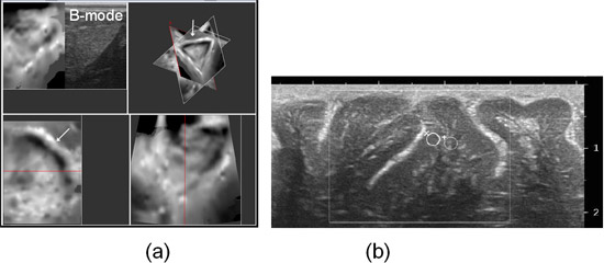
As well as conventional 2D elastography, palpation with a 3D transducer (55 mm x 60 mm surface) is frequently possible (Figure 3a). Early work has also begun on evaluating the application of shear wave elastography (SWE) in this area, using the Supersonic Imagine Aixplorer™, which allows measurement of Young’s modulus (YM) based on shear wave speed. YM estimates were obtained from regions of interest placed in grey and white matter (Figure 3b). Preliminary measurements suggest that grey matter has a YM that is greater (by about a factor of two) than that of white matter, which is contrary to some previous reports using other measurement methods.
 Fig. 3. (a) 3D intraoperative scan (orthogonal views) of a meningioma with a mobile boundary: the white region with underlying black at the boundary (arrows) indicates slip during palpation, and a clear dissection plane. (b) High resolution intraoperative image of brain cortical structure showing positioning of Young’s modulus measurement regions of interest over grey (x) and white (+) matter.