High resolution and microscopic elastography
E Elyas, L Garcia, JC Bamber, in collaboration with A Grimwood, Q Pankhurst, The Royal Institution and University College London; JD Holmes, G McKenzie, A Gilks, Michelson Diagnostics Ltd; Y Jamin, J Boult, S Robinson, CRUK-EPSRC Cancer Imaging Centre; R Sinkus, Laboratoire Ondes et Acoustique, ESPCI, Paris, France; J Erler, T Cox, Section of Cell and Molecular Biology.
Source of funding: EPSRC, CRUK, Michelson Diagnostics Ltd
The mechanical properties of the microenvironment interact with cell signalling to influence cell behaviour. Cells also manipulate the mechanical properties of their environment. Increased tissue shear modulus, for example, is associated with invasiveness. We are therefore developing lack tools to study these phenomena at high resolution in 3D cell culture and in vivo.
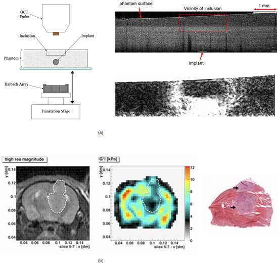
One approach uses optical coherence tomography (OCT), which provides optical backscatter cross-sectional images of tissue microanatomy, not dissimilar in character to ultrasound images, but with resolutions better than 10µm. We have been assessing the potential for imaging strains produced by non-contact actuation of a magnetisable implant. The field from a Halbach array was used to apply force to a 0.79 mm diameter steel sphere implanted in RTV silicone rubber of Young’s modulus (YM) 37k Pa containing optical scatterers and a rectangular inclusion of YM 269 kPa (Figure 7a). Good agreement was obtained between the strain fields predicted by finite element modelling and those measured using a 2D cross-correlation tracking algorithm to map the strains with a multifocus OCT microscope (EX 1301, Michelson Diagnostics). The sum of total shear strain and normal axial strain performed well at revealing the stiff inclusion (Figure 7a).
In another project, the dynamic magnetic resonance elastography technique of R Sinkus, ESPCI, Paris, has been implemented with assistance from R Sinkus on the 7 Tesla preclinical MRI scanner within the CRUK-EPSRC Imaging Centre. Preliminary results from an in vivo murine brain tumour, vibrating the intact skull at 1 kHz, show good correlation of reconstructions of the real part of the shear modulus with gross histology (Figure 7b).
 Fig. 7. Progress in microscopic and high resolution elastography. (a) OCT elastography of a rubber phantom (see text for description). (b) Preliminary preclinical magnetic resonance elastogram (storage shear modulus, cenre image) of an orthotopic murine RG2 glioma appearing softer than surrounding brain and showing good morphological correlation with histology (right). With thanks to Y Jamin, J Boult and S Robinson, Preclinical Biomarker Team, for the images in (b).