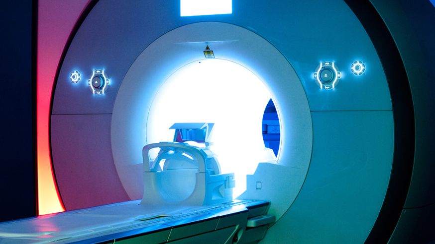
Magnetic resonance imaging (MRI) is able to detect indicators that a breast tumour is likely to be more aggressive, researchers have found.
The scans can identify the presence of fibrosis in a tumour – where connective tissue fibres are laid down around the cancerous cells, similar to scar tissue around an injury.
A high level of fibrosis is an indication that a breast tumour may be more aggressive, and is linked to a greater risk of the disease recurring and metastasising, as well as poorer patient survival.
Being able to detect these at-risk patients in a non-invasive way could allow clinicians to treat them sooner, potentially improving their prognosis.
Search is our twice-yearly newsletter to supporters. Read our latest news, recent research achievements and interviews with our world-leading scientists and clinicians.
Measuring tumour tissue for fibrosis
Writing in the journal European Radiology, the team from The Institute of Cancer Research, London, and The Royal Marsden NHS Foundation Trust explained how they performed their research, which was done in rats with chemically-induced breast tumours that show a range of fibrosis levels.
Using a clinical MRI scanner, the researchers imaged the rat tumours, taking a number of different measurements.
They then removed the tumours and used staining to look at how much collagen, the main protein that builds up in fibrosis, they contained, to determine which of the MRI measurements were sensitive to its presence.
The researchers found that a type of MRI called magnetisation transfer (MT) imaging, which is sensitive to structural integrity, was able to generate measurements that correlated well with the amount of collagen in the tumours – suggesting that MT imaging is able to identify fibrosis in breast cancer.
The research was supported by a range of organisations, including Cancer Research UK, the Engineering and Physical Sciences Research Council, the Medical Research Council and the Department of Health.
Study leader Dr Simon Robinson, Team Leader in Magnetic Resonance at the ICR, said:
“Our research shows that, using magnetisation transfer imaging, it is possible to detect fibrosis in breast cancers in a non-invasive way.
“Given that fibrosis is known to be a predictor of increased tumour aggressiveness and worse patient survival, the results of this study support the inclusion of MT imaging in clinical breast MRI examinations.”