A new radiotherapy technique could help doctors to focus treatment more precisely on tumours in the bladder and reduce damage to surrounding healthy tissue.
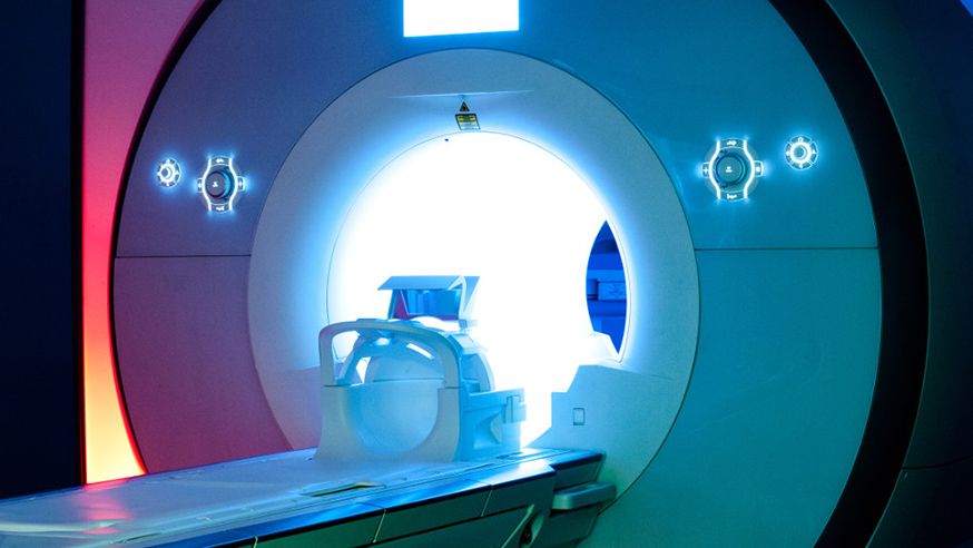
Researchers showed that pre-treatment imaging using magnetic resonance imaging (MRI) was effective at guiding radiotherapy towards tumours in the bladder.
Taking regular MRI scans of a tumour’s position helped to ensure that treatment to urinary bladder cancers could be delivered accurately during a course of radiotherapy.
Making sure that radiation hits the tumour site while avoiding healthy tissue is a key focus for radiotherapy research. Precision radiotherapy has seen huge advances in this area but challenges remain.
With bladder cancer, the area requiring treatment can shift depending on how much a patient drinks or how full the bladder is.
Researchers at The Institute of Cancer Research, London, The Royal Marsden NHS Foundation Trust and Aarhus University Hospital in Denmark used MRI scans to provide detailed pictures of bladder cancers and healthy soft tissue.
They used these images to adapt the radiotherapy dose before treatment, and then used scans to check the position of the tumour throughout the 10 minutes of treatment time that followed.
They found that the ‘adapted’ treatment successfully targeted the tumour throughout the course of treatment while minimising the impact on surrounding tissue.
Professor Uwe Oelfke, Head of the Joint Department of Physics at the ICR and The Royal Marsden, said: “Currently, patients are scanned before a course of radiotherapy to determine the location of the tumour in the bladder. Margins around the tumour are also treated to hopefully account for any tumour movement. It is less accurate than this new, improved method that recalculates and optimises treatment based on the images taken before treatment is delivered.
“This study shows that such re-optimised plans can accurately treat patients. In the future we expect that monitoring the treatment with MRI throughout a radiotherapy fraction on MRI-guided radiotherapy machines, such as our new MR Linac, will ensure accurate delivery and further reductions in safety margins.”
The researchers believe that this kind of approach – called MRI-guided adaptive radiotherapy (MRIGART) – will be a significant step forward in future radiotherapy treatment of patients with urinary bladder cancer.
The research also contributes towards optimising new state-of-the-art radiotherapy equipment, the MR Linac, that combines radiotherapy with a real-time MRI scanner. It is currently being built at the ICR’s and The Royal Marsden’s Sutton site and is set to revolutionise radiotherapy.
The study was published in the journal Radiotherapy and Oncology and was funded by the National Institute for Health Research Biomedical Research Centre for Cancer at The Royal Marsden and the ICR, Cancer Research UK and the CIRRO-Lundbeck Foundation.
