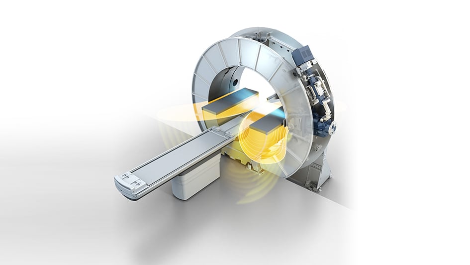
Image: MR Linac radiation beam and magnetic field combined illustration
Radiotherapy has come a long way since the early days of treating patients with cancer.
The theory behind radiotherapy is deceptively simple. Cancer is more prone to radiation than healthy cells in the body, so exposing them to radiation damages or kills cancer cells while leaving healthy cells relatively unscathed.
Radiotherapy is an integral part of cancer treatment. It plays a key role in alleviating symptoms and improving quality of life for cancer patients, and is involved in around 40 per cent of cancer cures.
Radiotherapy research is an important part of our research strategy here at The Institute of Cancer Research – as we seek to make treatment more precise and effective with lower rates of side effects.
Working closely with our hospital partner, The Royal Marsden, we are making improvements to treatment planning and delivery, and assessing innovative techniques to precisely target beams of radiation.
Both the technology of radiotherapy and the approaches taken to deliver it have advanced significantly in recent years, driven by innovative scientific and clinical research.
Some of the latest technological developments allow specialists to precisely target and destroy tumour cells, by adjusting a beam of radiation to the shape of tumours. These often involve using MRI or CT scans to accurately pinpoint tumour tissue while avoiding healthy cells – reducing side effects.
There are changes too in the way radiotherapy is given to patients, with a tendency to give fewer radiation treatments, each at a slightly increased dose, but with the effect of reducing the total cumulative dose.
Together these advances enable patients to maintain a higher quality of life during treatment, with fewer side effects and less time travelling to and from hospital.
The ICR works at the leading edge of imaging and precision radiotherapy. With state-of-the-art facilities and internationally renowned researchers, we are pioneering technologies to improve the diagnosis and monitoring of cancer, and to guide new forms of precision treatment.
Read more
Practice-changing trials
Here at the ICR, our researchers have pioneered radiotherapy research through the creation of new techniques and their evaluation in practice-changing clinical trials with The Royal Marsden.
In the 1980s, the ICR’s Professor Steve Webb applied mathematics to fine tune the shape of radiation beams, which helped in the development of intensity-modulated radiotherapy (IMRT) – a radiotherapy technique now used to treat many cancers.
IMRT enables the X-ray beam used in radiotherapy to be adjusted, both in shape and intensity, as the machine moves around the body. Altering the shape allows the beam to match the contours of the tumour, while changing the intensity enables higher doses of radiation to be concentrated on the cancerous cells, sparing the surrounding tissue.
Now a brand new imagining technology at the ICR and The Royal Marsden has demonstrated its power to shake up radiotherapy and change the role of all health professionals when delivering it to patients.
Pioneering the MR Linac
The MR Linac is a revolutionary piece of equipment which simultaneously generates magnetic resonance images and delivers X-ray beams for radiotherapy.
The ICR’s Professor Uwe Oelfke has been instrumental in developing and testing MRI to support treatment with the MR Linac. In September 2018, the ICR and The Royal Marsden delivered the first ever treatment in the UK using the MR Linac machine.
MRI takes highly detailed images of tissues inside the body using powerful electromagnets, so combining MRI and radiotherapy could help increase the accuracy of treatment and deliver better outcomes for patients.
Currently, patients with cancer might have an MRI in the days or weeks leading up to radiotherapy to help plan their treatment. But actions like breathing, eating or drinking can all cause our organs to expand or contract – and that in turn may change the position of the tumour.
Scans taken days or even hours before radiotherapy is delivered may not accurately reflect the situation at the time of treatment.
Using the MR Linac, scans can be taken at the same time as radiotherapy is delivered, so treatment can immediately be adjusted to give the most accurate doses of radiation to tumours.
Higher-definition images
Cutting-edge technology like the MR Linac is the latest example of how increasingly sophisticated imaging techniques are improving radiotherapy.
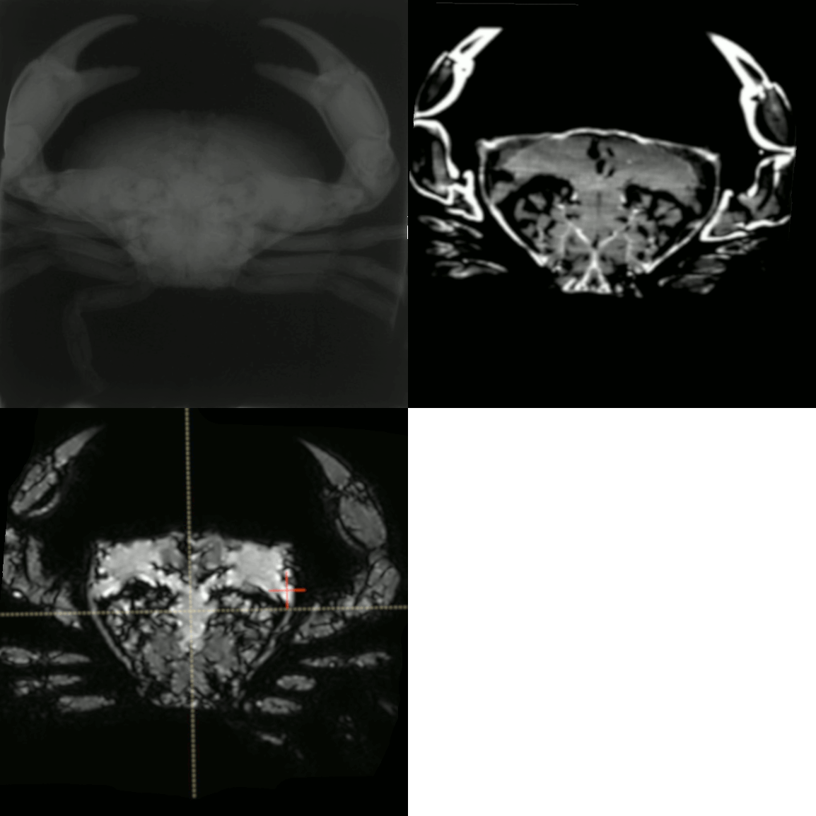
A paper published in the journal Radiography shows the massive increase in definition that can be seen using the MR Linac compared with older technologies:

Image: Animated GIF compiled using images from paper in Radiography journal
Using a crab as a test subject, the study highlights the increasing level of detail that can be seen – from CT scans that were used to plan radiotherapy in the 1980s and 1990s to those used in treatment today, and finally to MRI scans taken at the same time as radiotherapy using the MR Linac.
Changing role of radiographers
Technology is moving quickly in this field, so it’s not just important to train more staff – but to train them in the right things. We need to make sure that training keeps up with the latest research, so that radiographers learn the very latest technologies and techniques.
Advances in technology like the MR Linac could revolutionise radiotherapy, but to deliver on their promise we need more clinicians with expertise in both MRI and radiotherapy.
Having skilled – and research active – clinical oncologists, physicists, radiographers, allied healthcare staff and maintenance crew will be vital to create the right environment to promote innovation in the clinic, and ultimately improve outcomes for patients.
And the ICR and The Royal Marsden we are doing just that with the MR Linac. We are training radiographers in the science of MR imaging, so they can adjust radiotherapy based on MRI scans taken just before their treatment.
Delivering radiotherapy to cancer patients
Dr Helen McNair is a radiographer at The Royal Marsden and a member of the ICR’s Honorary Faculty. In 2018 she was named UK Radiographer of the Year by The Royal Society of Radiographers.
Therapeutic radiographers like Dr McNair are responsible for the delivery of radiotherapy for cancer patients. Radiography is both a caring and a technical role, supporting patients through every step of radiotherapy while optimising their treatment using a wide range of technologies.
Dr McNair has played a pivotal role in supervising research and mentoring radiographers at the ICR and The Royal Marsden.
Radiographers have a wealth of knowledge built up through their daily interactions with patients, and as technology like the MR Linac increases the capabilities of radiotherapy, it makes sense to use their expertise to plan better treatment.
Currently, radiographers, physicists and clinical oncologists are all required to be present at the time of treatment, which can take up to 45 minutes. This is very resource intensive.
Ideally, radiographers trained to use the MR Linac would able to scan patients when they arrive for treatment, check their treatment plans are correct along with a physicist, and deliver radiotherapy all in one sitting. But we aren’t there yet.
With additional training radiographers could make routine adjustments to the treatment plan without the need to consult the oncologist, so they can get on with helping other patients.
Improving care
Dr McNair has just received a £600,000 grant from the NIHR to investigate how to make this process of real-time adaptive radiotherapy possible.
Training more radiographers to interpret MRI scans is also key to ensuring this technology can be used more widely, and the ICR and The Royal Marsden together organise regular training courses to equip radiographers with the tools they need to understand MRI scans.
Dr McNair says: “With new radiotherapy technologies and treatment strategies improving care it’s an exciting time to be working in the field. I feel proud of the research I have been involved with to improve radiotherapy for patients.
“We are making real advances with radiotherapy research, but taking these from bench to bedside quickly will only be possible with collaborative teams of multidisciplinary experts.
“Therapeutic radiographers and the many different highly skilled staff who play a role in the health service are essential to delivering world-class radiotherapy for the 21st century.”
New imaging technologies have already had a dramatic impact on how radiotherapy is delivered for patients – and the best, it seems, is yet to come.
comments powered by