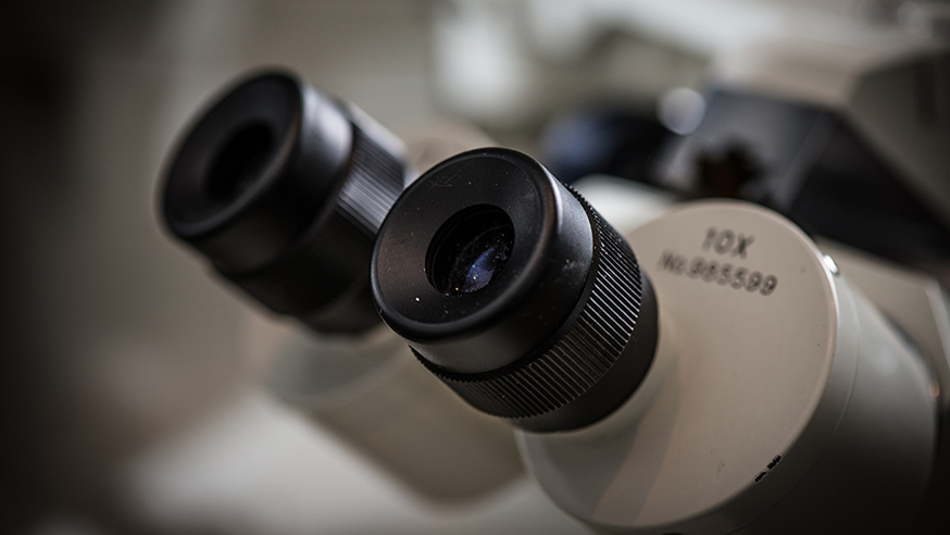
Image: Liquid helium cryogenic transmission electron microscope (Credit: EMSL/CC BY-NC-SA 2.0)
Nobel prizes can shine a light on areas of science which are making a huge impact in laboratories across the world, but may not be well known among the wider public.
This week, many of our researchers are excited by the news that the pioneers of a technology called cryo-electron microscopy – cryo-EM for short – have won the 2017 Nobel Prize for Chemistry.
Cryo-EM – which involves freezing samples to -180oC – allows researchers to visualise samples in such fine detail that they can see the structure of their atoms. It’s a technique which is being put to good use here at The Institute of Cancer Research in London for understanding cancer on a molecular level.
The winners of the prize led research that has given cancer scientists a brand new tool to use in understanding the very fundamental workings of cancer, and which could lead to new drugs.
Revolutionary technology
Dr Ed Morris, Team Leader in Structural Electron Microscopy at the ICR, is an enthusiastic proponent of the power of Cryo-EM. He said:
“Cryo-electron microscopy is revolutionising molecular and cellular biology, allowing the detailed understanding of the molecular machines within cells and how they work. This type of information is crucial, for example, in identifying new therapeutic targets and in designing drugs to treat diseases such as cancer.
Dr Morris added: “It is wonderful to hear the news that Jacques Dubochet, Joachim Frank and Richard Henderson have been awarded the 2017 Nobel Prize in Chemistry for their work.
“They each richly deserve the award, a prestigious and extremely timely recognition of an area of scientific research which is already transforming the way we look at the inner workings of cells.”
Cryo-electron microscopy in cancer research
Alongside colleagues at the Medical Research Council Laboratory of Molecular Biology in Cambridge – where Professor Henderson made his discoveries – Dr Morris’s team has used cryo-EM to make images of some of the proteins that drive cell division.
These proteins are not only often malfunctioning in cancer, but are also involved in some of the most important processes in life – overseeing the production of new cells, and the building of whole organisms from single cells.
Partnership
“Cryo-electron microscopy is widely used within the Division of Structural Biology at ICR to study a range of protein complexes,” said Dr Morris.
“This includes the proteasome, a cellular recycling unit with a role in many diseases, where cryo-electron microscopy was used to view a drug related inhibitor bound to its active sites. We are also investing in the latest cryo-electron microscope technology as part of a central London consortium, which will allow us to increase significantly our capabilities in this increasingly important and exciting field.”
This consortium was awarded £3m this year from the Wellcome Trust, which includes the purchase of a new cryo-EM machine called the Krios and which will strengthen existing research partnerships at the joint ICR and Imperial Centre for Cancer Research Excellence.
Responding to the launch of this programme, Professor Jon Pines, the ICR’s Head of Cancer Biology, explained the benefits of the technology for cancer research.
“It not only offers far higher resolution than traditional electron microscopy methods – allowing the visualisation of atomic structures – but also means we can study protein complexes in conditions closer to those in the human body,” he said. “Cryo-EM should make it much easier to design new, more potent cancer drugs.”
Basic science
The Nobel Prize will increase the profile of cryo-electron microscopy – and helps to underline the importance of investment in early, basic science that can lead to benefits years later.
As our researchers at the ICR and our partners use the technology to gain new understandings of cancer’s fundamental biology, they will discover weaknesses that could be targeted by the next generation of drugs.
comments powered by