At The Institute of Cancer Research here in London, we are always looking out for new and exciting ways to communicate about our research, from videos and infographics to interactive displays and late-night museum sessions.
We set our researchers the challenge this year of capturing in a single image the groundbreaking work they are doing to make the discoveries that defeat cancer.
The response we had was fantastic! Our researchers really pulled out the stops to submit breathtaking and original images that showcase the vital research we conduct at the ICR.
Here are some of our favourite entries:
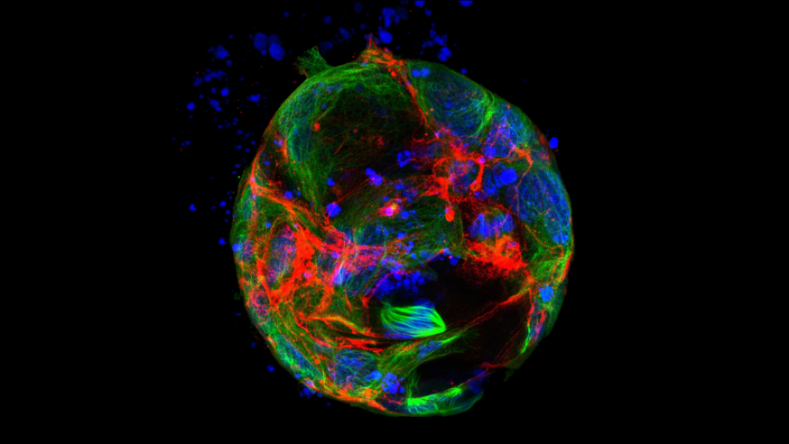
Winner: Dr Gianmaria Liccardi, Division of Breast Cancer Research

Our winning image highlights in vivid detail how loss of RIPK1 – a key signalling molecule – causes cells’ chromosomes to become unstable and degrade (green).
Dr Liccardi switched off the RIPK1 gene in intestinal cells and used a technique called fluorescence microscopy to monitor the impact this had on the function of the cells.
Studies such as these are an important aspect of our research as they enable us to understand more about the biology of cancer and offer potential new clues for targeting tumour cells.
Shortlisted: Mateus Crespo, Division of Clinical Studies

One of the biggest challenges our researchers face when they are developing new drugs is overcoming resistance.
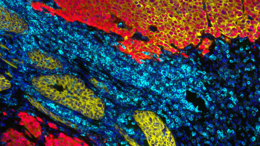
Mateus has beautifully captured the development of resistance to the drug abiraterone in a patient with prostate cancer.
Abiraterone – which was discovered here at the ICR – is used to treat men with advanced prostate cancer, and is benefiting hundreds of thousands of patients worldwide.
But mutations to the androgen receptor gene can allow cancer cells to become resistant to abiraterone. This is shown in the image by the maintained expression of the androgen receptor (red) following treatment with abiraterone.
Shortlisted: Valeria Molinari, Louise Howell, Dr Maria Vinci, Katy Taylor and Professor Chris Jones, Division of Molecular Pathology

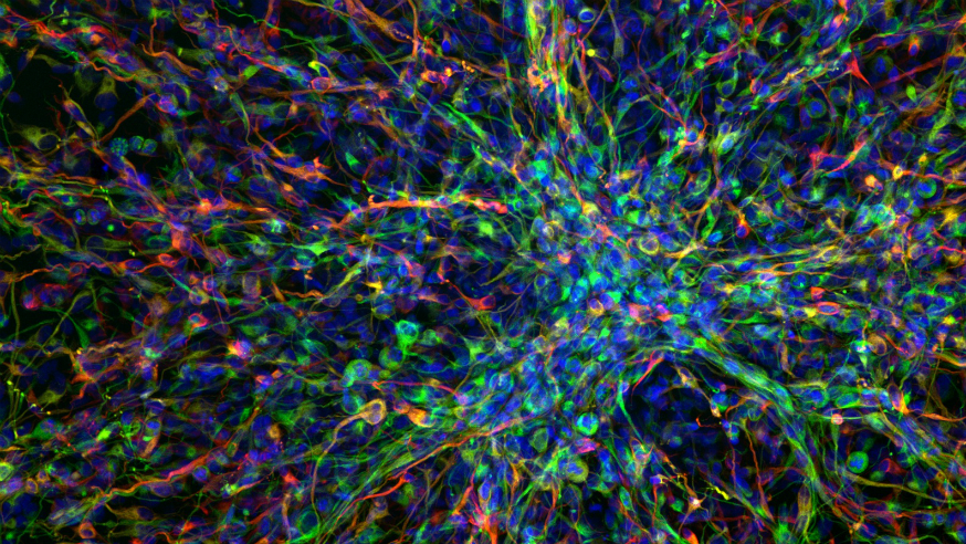
Glioblastoma multiforme is a highly aggressive and difficult-to-treat childhood brain tumour with an average survival for patients of 12–15 months.
In this image, the team embedded a 10-year-old patient’s tumour cells in a gel-like matrix and allowed the cells to spread, mimicking the pattern of tumour growth that occurs in the brain. They were able to visualise this movement using a type of microscopy called confocal microscopy.
The researchers hope that studies like this, which try to better represent conditions found in the body, will increase our understanding of the biology of this devastating disease, and ultimately lead to the development of new treatments.
This year’s entries have certainly set the bar high – we can’t wait to see how our researchers will rise to the challenge in 2016!
comments powered by